Материал: Kaplan USMLE-1 (2013) - Anatomy

Внимание! Если размещение файла нарушает Ваши авторские права, то обязательно сообщите нам

Chapter 3 • Abdomen, Pelvis, and Perineum

Extrahepatic BiliaryAtresia

Occurs when the lumen ofthe biliary ducts is occluded owing to incomplete re­ canalization.This condition is associated with jaundice, white-colored stool, and dark-colored urine.

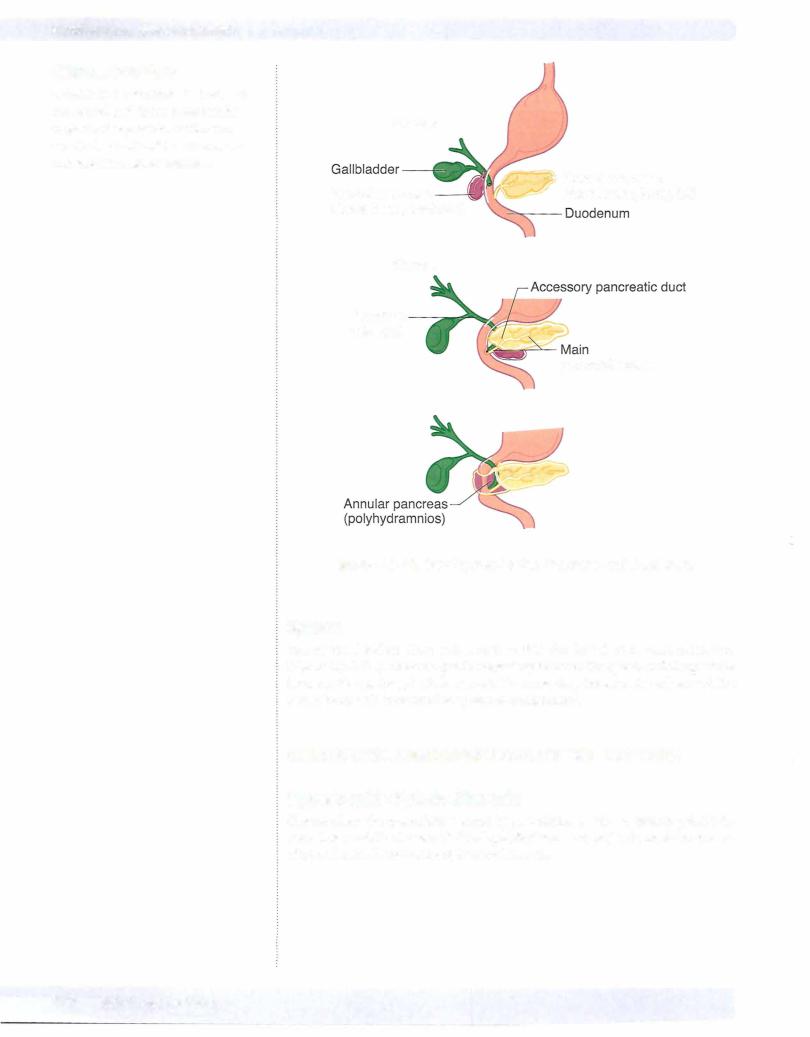
Annular Pancreas

Occurs when the ventral and dorsal pancreatic buds form a ring around the duo­ denum, thereby causing an obstruction ofthe duodenum and polyhydrarnnios

DuodenalAtresia

Occurs when the lumen ofthe duodenum is occluded owing to failed recanaliza­ tion. This condition is associated with polyhydramnios, bile-containing vomitus, and a distended stomach.

Omphalocele

An omphalocele occurs when the midgut loop fails to return to the abdominal cavity and remains in the umbilical stalk.

•The viscera herniate through the umbilical ring and are contained in a shinysac of amnion at the base of the umbilical cord.

•Omphalocele is often associated with multiple anomalies of the heart and nervous system with a high mortality rate (25%).

Gastroschisis

Gastroschisis occurs when the abdominal viscera herniate through the body wall directly into the amniotic cavity, usually to the right of the umbilicus.

•This is a defect in the closure of the lateral body folds and a weakness of the anterior wall at the site of absorption of the right umbilical vein.

•Note that the viscera do not protrude through the umbilical ring and are not enclosed in a sac of amnion.

lleal (Meckel) Diverticulum

Occurs when a remnant of the vitelline duct persists, thereby forming a blind pouch on the antimesenteric border of the ileum. This condition is often asymp­ tomatic but occasionally becomes inflamed ifit contains ectopic gastric, pancre­ atic, or endometrial tissue, which may produce ulceration. They are found 2 feet from the ileocecal junction, are 2 inches long, and appear in 2% ofthe population.

Vitelline Fistula

Occurs when the vitelline duct persists, thereby forming a direct connection be­ tween the intestinal lumen and the outside ofthebodyatthe umbilicus. This condi­ tion is associated with drainage ofmeconium from the umbilicus.
MEDICAL 249

Section Ill • Gross Anatomy

Malrotation of Midgut

Occurs when the midgut undergoes only partial rotation and results in abnormal position of abdominal viscera. This condition may be associated with volvulus (twisting of intestines) .

Colonic Aganglionosis (Hirschsprung Disease)

Results from the failure ofneural crest cells to form the myenteric plexus in the sigmoid colon and rectum. This condition is associated with loss of peristalsis and immobility of the hindgut, fecal retention and abdominal distention of the transverse colon (megacolon).

ABDOMINALVISCERA

Liver

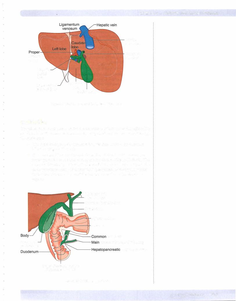
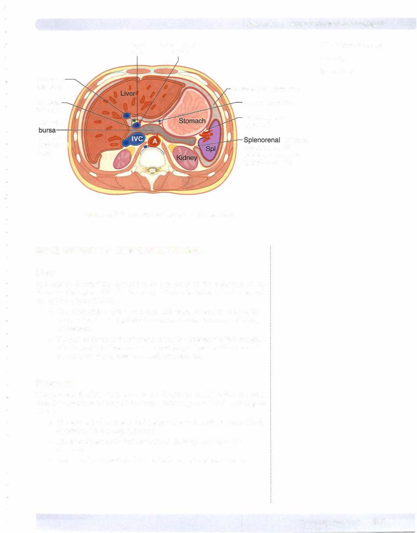
The liver has 2 surfaces: a superior or a visceral surface (Figure III-3- 1 abdominal cavity and is protected by peritoneum:

or diaphragmatic surface and an inferior 1) . lt lies mostly in the right aspect of the the rib cage. The liver is invested by visceral

•The reflection of visceral peritoneum between the diaphragmatic surface of the liver and the diaphragm forms the falciform ligament, which continues onto the liver as the coronary ligament and the right and left triangular ligaments.

•The extension of visceral peritoneum between the visceral surface of the liver and the first part of the duodenum and the lesser curvature of the stomach forms the hepatoduodenal and hepatogastric ligaments of the lesser omentum, respectively.

The liver is divided into 2 lobes of unequal size as described below (Figure Ill-

3- 1 1).

•Fissures for the ligamentum teres and the ligamentum venosum, the porta hepatis, and the fossa for the gallbladder further subdivide the right lobe into the right lobe proper, the quadrate lobe, and the caudate lobe.

•The quadrate and caudate lobes are anatomically part of the right lobe but functionally part of the left. They receive their blood supply from the left branches of the portal vein and hepatic artery and secrete bile to the left hepatic duct.

The liver has a central hilus, or porta hepatis, which receives venous blood from the portal vein and arterial blood from the hepatic artery.

•The central hilus also transmits the common bile duct, which collects bile produced by the liver.

•These structures, known collectively as the portal triad, are located in the hepatoduodenal ligament, which is the right free border of the lesser omentum.

The hepatic veins drain the liver by collecting blood from the liver sinusoids and returning it to the inferior vena cava.

250 MEDICAL

Источник: https://studfile.net/preview/14638320/