Section IV • Neuroscience
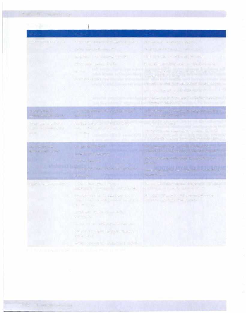
Table IV-1-1
Disease |
Symptoms |
Multiple sclerosis (MS) |
Symptoms separated in space and time |
|
Vision loss (optic neuritis) |
|
Internuclear ophthalmoplegia (MLF) |
|
Motor and sensory deficits |
|
Vertigo |
|
Neuropsychiatric |
Metachromatic |
Four types; motor and cognitive issues |
leukodystrophy (MLD) |
with seizures |
Progressive multifocal |
Cortical myelin commonly affected; |
leukoencephalopathy |
causes limb weakness, speech |
(PML) |
problems |
Central pontine |
Pseudobulbar palsy |
myelinolysis (CPM) |
Spastic quadriparesis |
|
|
Mental changes |
|
May produce the "locked-in" syndrome |
|
Often fatal |
Guillain-Barre syndrome |
Acute symmetric ascending |
|
inflammatory neuropathy of PNS myelin |
|
Weakness begins in lower limbs and |
|
ascends; respiratory failure can occur in |
|
severe cases |
|
Autonomic dysfunction may be |
|
prominent |
|
Cranial nerve involvement is common |
|
Sensory loss, pain, and paresthesias |
|
rarely occur |
|
Reflexes invariably decreased or absent |
Abbreviations: CJD, Jacob-Creutzfeldt; MLF, medial longitudinal fasciculus
Notes
Occurs twice as often in women
Onset often in third or fourth decade
Higher prevalence in temperate zones
Relapsing-remitting course is most common
Well-circumscribed demyelinated plaques often in periventricular areas
Chronic inflammation; axons initially preserved
Increased lgG (oligoclonal bands) in CSF
Treatment: high-dose steroids, interferon-beta, glatiramer (Copaxone®)
Arylsulfatase A deficiency in lysosomes affects botn CNS and PNS myelin
Caused by CJD virus
Affects immunocompromised, especially AIDS
Demyelination, astrogliosis, lymphohistiocytosis
Focal demyelination of central area of basis pontis (affects corticospina corticobulbartracts)
Seen in severely malnourished, alcoholics, liver disease
Probably caused by overly aggressive correction of hyponatremia
Two-thirds of patients have history of respiratory or GI illness 1-3 weeks prior to onset
Elevated CSF protein with normal cell count (alburninocytologic dissociation)