Chapter 1 • NervousSystem
ChapterSummary
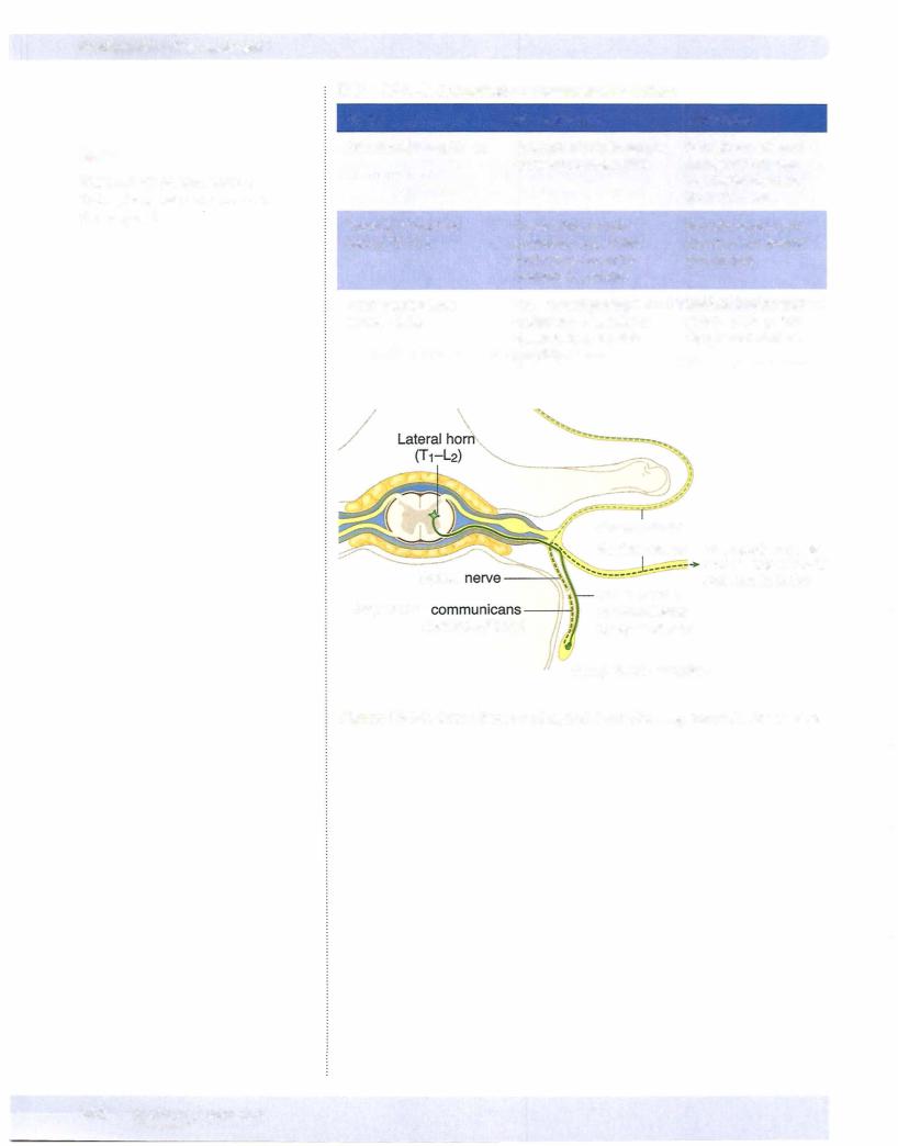
•The peripheral nervous system (PNS) consists of 1 2 pairs of cranial nerves, 31 pairs of spinal nerves with their related sensory and motor ganglia, and the peripheral part ofthe autonomic nervous system. The afferent and efferent neurons in the PNS convey somatic and visceral (autonomic) functions to and from the central nervous system (CNS).
Cells of Nervous System
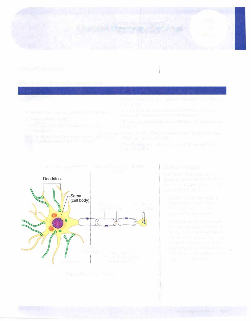
•The basic functional cell for conducting motor and sensory functions within the nervous system is the neuron. Neurons in the CNS are myelinated by oligodendrocytes, and in the PNS neurons are myelinated by Schwann cells. Oligodendrocytes myelinate multiple axons but Schwann cells myelinate only a segment of one axon.
•The skeletal motor neurons and preganglionic motor neurons in the CNS develop from the neural tube, whereas the sensory neurons and postganglionic neurons located in sensory or autonomic ganglia, respectively, in the PNS derive from neural crest cells.
•Neurulation and the development of the nervous system begin in the third week of development. As the primitive streak regresses caudally, the notochord develops in the midaxis ofthe embryo between the
buccopharyngeal membrane and the cloaca[ membrane. The appearance of the notochord then induces the ectoderm overlying the notochord to form the neural plate composed of neuroectoderm. The neural plate is wide at the cranial end and tapers caudally. By the end of the third week, the lateral margins of the neural plate thicken and become elevated to form the neural folds with the neural groove located centrally between the 2 folds. The neural folds then grow over the midline and begin to fuse to form the neural tube. Closure ofthe neural tube begins in the cervical region and continues cranially and caudally. The cephalic (cranial neuropore) and the caudal (caudal neuropore) ends ofthe neural tube close last. Failure of closure of the cranial and caudal neuropores results in anencephaly and spina bifida. Alpha-fetoprotein levels are increased with the neural tube defects. During closure of the neural tube, neural crest cells are formed from neuroectoderm at the margins of the neural folds. The neural crest cells migrate throughout the embryo and form a number of cell types.