CSF DISTRIBUTION, SECRETION, AND CIRCULATION
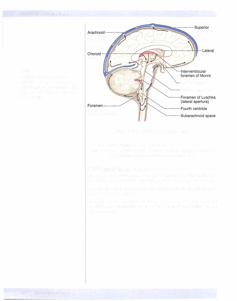
CSF fills the subarachnoid space and the ventricles ofthe brain. The average adult has 90 to 1 50 mL of total CSF, although 400 to 500 mL is produced daily. Only 25 mL of CSF is found in the ventricles themselves.
Approximately 70% of the CSF is secreted by the choroid plexus, which consists of glomerular tufts of capillaries covered by ependymal cells that project into the ventricles (the remaining 30% represents metabolic water production). The cho roid plexus is located in parts ofeach lateral ventricle, the third ventricle, and the fourth ventricle.
CSF from the lateral ventricles passes through the interventricular foramina of Monro into the third ventricle. From there, CSF flows through the aqueduct of Sylvius into the fourth ventricle. The only sites where CSF can leave the ventricles and enter the subarachnoid space outside the CNS are through 3 openings in the fourth ventricle, 2 lateral foramina of Luschka and the median foramen of Magendie.
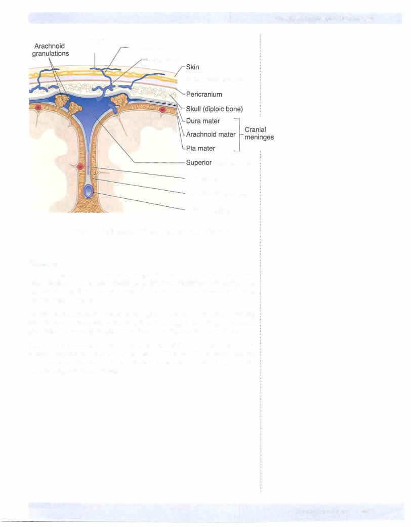
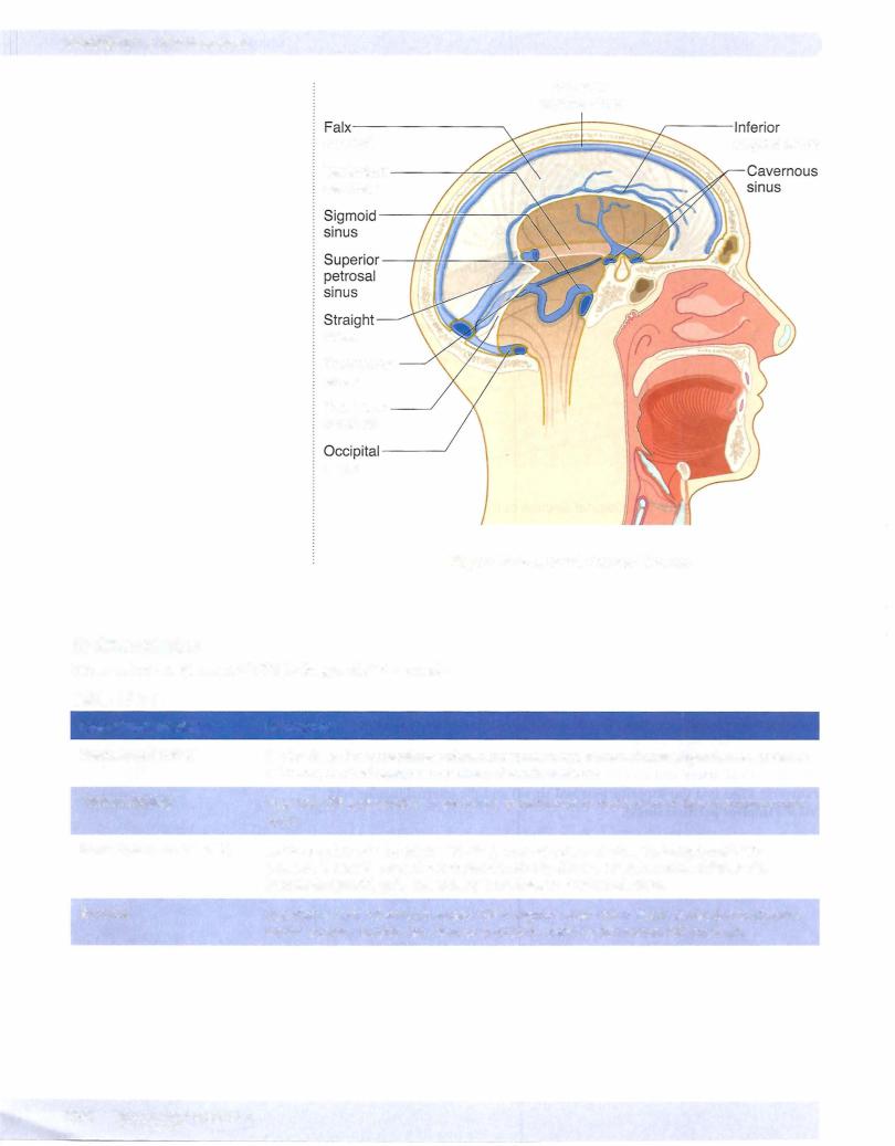
Within the subarachnoid space, CSF also flows up over the convexity ofthe brain and around the spinal cord. Almost allCSF returns to the venous system by drain ing through arachnoid granulations into the superior sagittal dural venous sinus.
Normal CSF is a clearfluid, isotonic with serum (290-295 mOsm/L).
The pH of CSF is 7.33 (arterial blood pH, 7.40; venous blood pH, 7.36).
Sodium ion (Na+) concentration is equal in serum and CSF (:o:l38 mEq/L).
CSF has a higher concentration of chloride (CI-) and magnesium (Mg2+) ions than does serum.
CSF has a lower concentration ofpotassium (K+), calcium (Ca2+), and bicarbon ate (HC03) ions, as well as glucose, than does serum.
The Blood-Brain Barrier and the Blood-CSF Barrier
The chemical integrity of the brain is protected in a different way by 2 separate systems.
Chapter 3 • Ventricular System
Clinical Correlate
The concentration of protein (including all immunoglobulins) is much lower in the CSF as compared with serum.
Normal CSFcontains 0 to 4 lymphocytes or mononuclear cells per cubic millimeter. Although the presence ofa few monocytes or lymphocytes is normal, the presence of polymorphonuclear leukocytes
is always abnormal, as in bacterial meningitis.
Red blood cells (RBCs) are not normally found in the CSF but may be present after traumatic spinal tap or subarachnoid hemorrhage.
Increased protein levels may indicate a CNS tumor.
Tumor cells may be present in the CSF in cases with meningeal involvement.