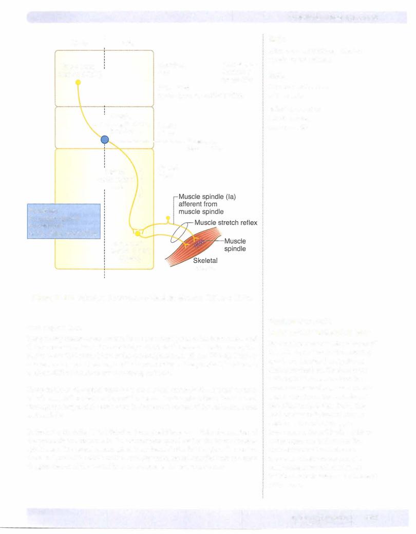
Both ends ofthe muscle spindle are connected in parallel with the extrafusal fibers, so these receptors monitor the length and rate of change in length of extrafusal fibers. Muscles involved with fine movements contain a greater density of spindles than those used in coarse movements.
Commonlytested muscle stretch reflex.es
The deep tendon (stretch, myotatic) reflex is monosynaptic and ipsilateral. The afferent limb consists of a muscle spindle, Ia sensory neuron, and efferent limb
(lower motor neuron). These reflexes are useful in the clinical exam.
Reflex |
Cord Segment Involved |
Muscle Tested |
Knee (patellar) |
L2-L4 (femoral n.) |
Quadriceps |
Ankle |
51 (tibial n.) |
Gastrocnemius |
Elbow |
C5-C6 |
(musculocutaneous n.) |
Biceps |
Elbow |
C7-C8 |
(radial n.) |
Triceps |
Forearm |
C5-C6 |
(radial n.) |
Brachioradialis |
Muscle stretch (myotatic) reflex
1be muscle stretch (myotatic) reflex is the stereotyped contraction of a muscle in response to stretch of that muscle. The stretch reflex is a basic reflexthat oc curs in allmuscles and is the primary mechanism for regulating muscle tone.
Muscle tone is the tension present in all resting muscle. Tension is controlled by the stretch reflexes.
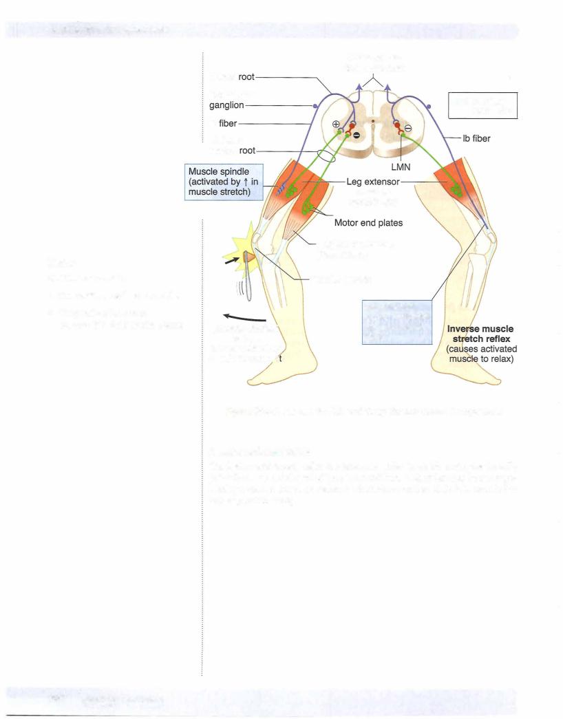
The best example of a muscle stretch or deep tendon reflex is the knee-jerk re flex. Tapping the patellar ligament stretches the quadriceps muscle and its muscle spindles. Stretch of the spindles activates sensory endings (Ia afferents), and af ferent impulses are transmitted to the cord. Some impulses from stretch recep tors carried by Ia fibers monosynaptically stimulate the alpha motoneurons that supply the quadriceps. This causes contraction ofthe muscle and a sudden exten sion of the leg at the knee. Afferent impulses simultaneously inhibit antagonist muscles through interneurons (in this case, hamstrings).
Inverse muscle stretch reflex
The inverse muscle stretch reflex monitors muscle tension. This reflex uses Golgi tendon organs (GTOs). These are encapsulated groups of nerve endings that ter minate between collagenous tendon fibers at the junction of muscle and tendon. GTOs are oriented in series with the extrafusal fibers and respond to increases in force or tension generated in that muscle. Increases in force in a muscle increase the firing rate of lb afferent neurons that innervate the GTOs, which, in turn, polysynaptically facilitate antagonists and inhibit agonist muscles.
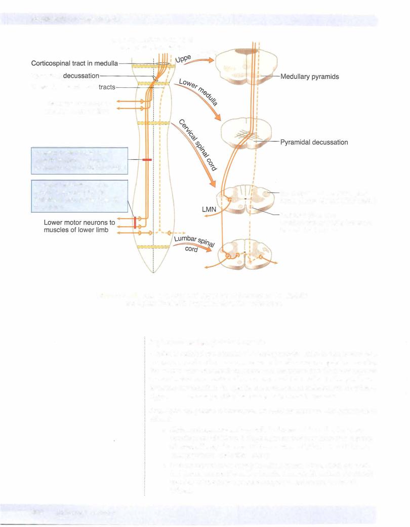
Muscle tone and reflex activity can be influenced by gamma motoneurons and by upper motoneurons. Gamma motoneurons directly innervate the muscle spindles and regulate their sensitivity to stretch. Upper motoneurons innervate gamma motoneurons and also influence the sensitivity of muscle spindles to stretch. Stimulation of gamma motoneurons causes intrafusal muscle fibers lo cated at the pole of each muscle spindle to contract, which activates alpha moto neurons, causing an increase in muscle tone.