Chapter If • The Spinal Cord
Clinical Correlate
Upper Motoneuron Versus Lower Motoneuron Muscle Lesions
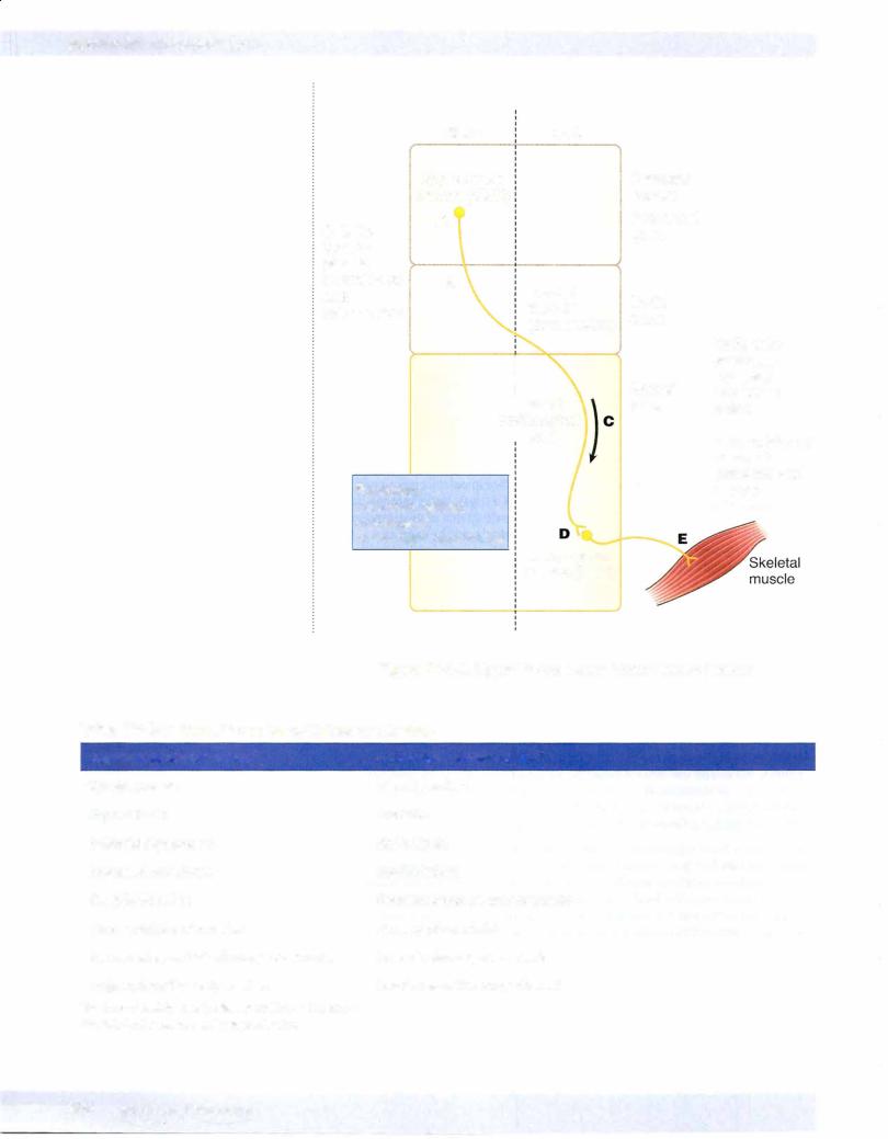
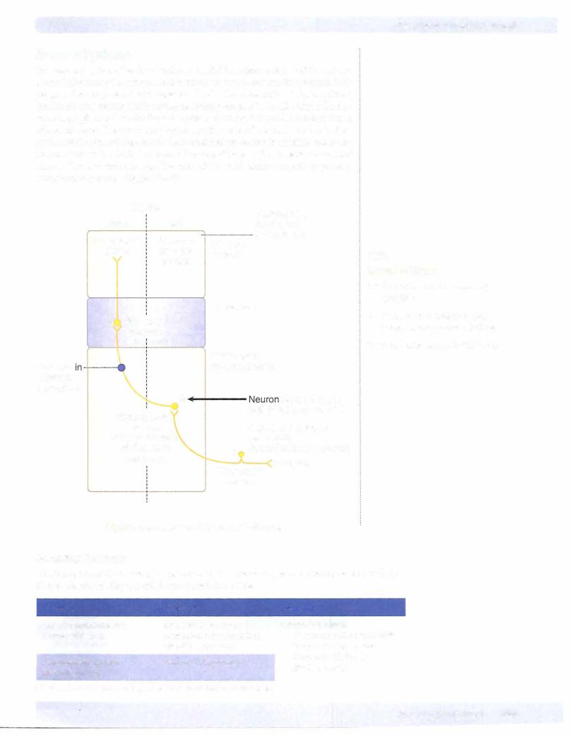
A fundamental requirement of interpreting the cause of motor weakness in neuroscience cases is the ability to distinguish between a lesion of an upper versus a lower motoneuron. Because a lesion to either an upper or a lower motoneuron produces a weakness in the ability to voluntarily contract skeletal muscles, the key to distinguishing an upper from a lower motoneuron lesion will be the condition of reflexes ofthe affected muscles (Figure IV-4-5).
A lesion of any part of a lower motoneuron will result in hypoactive muscle stretch reflexes and a reduction in muscle tone (hypotonicity) because lower motoneurons form the motor component of the reflex. Therefore, lower motoneuron lesions result in a paresis combined with suppressed or absent muscle stretch reflexes. An early sign of a lower motoneuron lesion is muscle fasciculations, which are twitches or contractions of groups of muscle fibers, that may produce a twitch visible on the skin. Later, lower motoneuron lesions produce fibrillations, which are invisible 1- to 5-ms potentials, detected with electromyography. Muscles denervated by a lower motoneuron lesion undergo pronounced wasting or atrophy. The constellation of lower motoneuron lesion signs combining paresis with suppressed or absent reflexes, fasciculations, and atrophy is known as a flaccid paralysis. With few exceptions, lower motoneuron lesions produce a flaccid paralysis ipsilateral and at the level of the lesion.
Neurologically, upper motoneurons includingthe corticospinal tract have a net overall inhibitory effect on muscle stretch reflexes. As a result, upper motoneuron lesions combine paresis ofskeletal muscles with muscle stretch or deep tendon reflexes that are hyperactive or hypertonic. The hypertonia may be seen as decorticate rigidity (i.e., postural flexion ofthe arm and extension ofthe leg) or decerebrate rigidity (i.e., postural extension ofthe arm and leg) depending on the location ofthe lesion. Lesions above the midbrain produce decorticate rigidity; lesions below the midbrain produce decerebrate rigidity. Upper motoneuron lesions result in atrophy ofweakened muscles only as a result ofdisuse, because these muscles can still be contracted by stimulating muscle stretch reflexes.
Upper motoneuron lesions are also accompanied by reversal of cutaneous reflexes, which normally yield a flexor motor response. The best known of the altered flexor reflexes is the Babinski sign. The test for the Babinski reflex is performed by stroking the lateral surface of the sole ofthe foot with a slightly painful stimulus. Normally, there is plantar flexion of the big toe. With a lesion ofthe corticospinal tract, the Babinski reflex is present, which is characterized by extension of the great toe and fanning of the other toes. Two other flexor reflexes, the abdominal and cremasteric, are also lost in upper motoneuron lesions. The constellation of upper motoneuron lesion signs combining paresis with increases or hyperactive reflexes, disuse atrophy of skeletal muscles, and altered cutaneous reflexes is known as a spastic paresis.
In contrastto lower motoneuron lesions, lesions of upper motoneurons result in a spastic paresis that is ipsilateral or contralateral and below the site of the lesion. Upper motoneuron lesions anywhere in the spinal cord will result in an ipsilateral spastic paresis below the level of the lesion. Upper motoneuron lesions between the cerebral cortex and the medulla above the decussation ofthe pyramids will result in a contralateral spastic paresis below the level of the lesion.