Материал: Kaplan USMLE-1 (2013) - Anatomy

Внимание! Если размещение файла нарушает Ваши авторские права, то обязательно сообщите нам

Section IV • Neuroscience

Clinical Correlate

Tabes patients present with paresthesias (pins-and-needles sensations), pain, polyuria, Romberg sign.

Clinical Correlate

Spastic bladder results from lesions of the spinal cord above the sacral spinal cord levels. There is a loss of inhibition of the parasympathetic nerve fibers that innervate the detrusor muscle during the filling stage. Thus, the detrusor muscle responds to a minimum amount of stretch, causing urge incontinence.

Clinical Correlate

Atonic bladder results from lesions to the sacral spinal cord segments or the sacral spinal nerve roots. Loss of pelvic splanchnic motor innervation

with loss of contraction of the detrusor muscle results in a full bladder with a continuous dribble of urine from the bladder.

378 MEDICAL

Brown-Sequardsyndrome

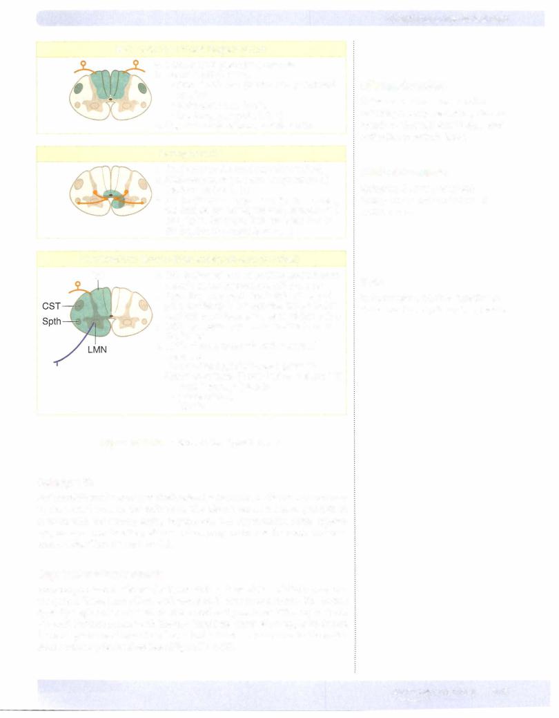
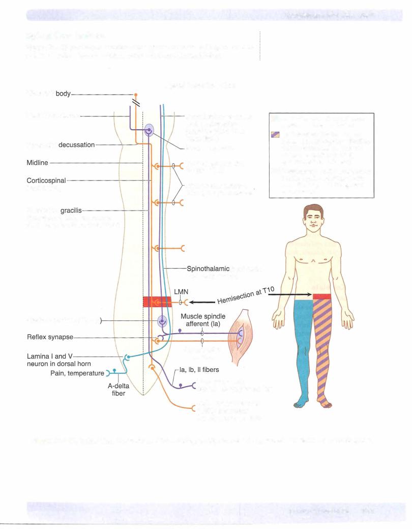
Hemisection of the cord results in a lesion of each of the 3 main neural systems: the principal upper motoneuron pathway of the corticospinal tract, one or both dorsal columns, and the spinothalamic tract. The hallmark of a lesion to these 3 long tracts is that the patient presents with 2 ipsilateral signs and one contralat­ eral sign. Lesion of the corticospinal tract results in an ipsilateral spastic paresis below the level of the injury. Lesion to the fasciculus gracilis or cuneatus results in an ipsilateral loss ofjoint position sense, tactile discrimination, and vibratory sensations below the lesion. Lesion ofthe spinothalamic tract results in a contra­ lateral loss of pain and temperature sensation starting one or 2 segments below the level of the lesion. At the level of the lesion, there willbe an ipsilateral loss of allsensation, including touch modalities as well as pain and temperature, and an ipsilateral flaccid paralysis in muscles supplied by the injured spinal cord seg­ ments (Figure IV-4-15).

Polio

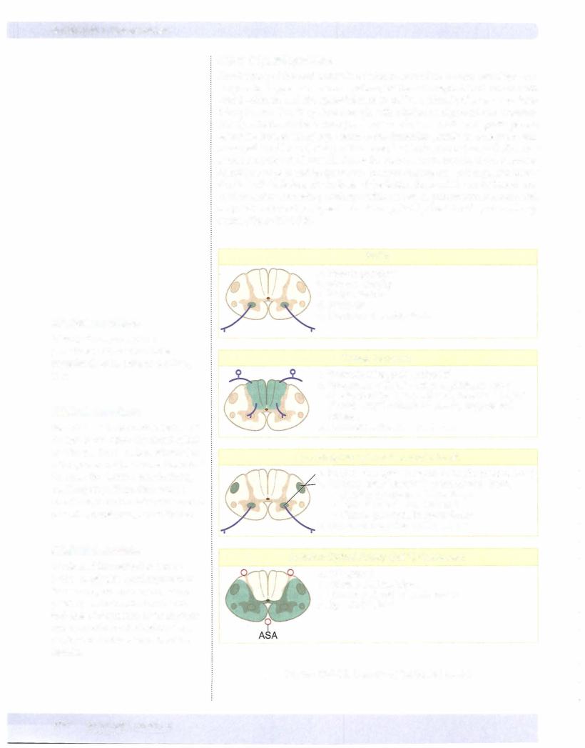
a.Flaccid paralysis

b.Muscle atrophy

c.Fasciculations

d.Areflexia

e.Common at lumbar levels

Tabes Dorsalis

a."Paresthesias, pain, polyuria"

b.Associated with late-stage syphilis, sensory ataxia, positive Romberg sign: sways with eyes closed, Argyll Robertson pupils, suppressed reflexes

c.Common at lumbar cord levels

Amyotrophic Lateral Sclerosis (ALS)

a.Progressive spinal muscular atrophy (ventral horn)

b.Primary lateral sclerosis (corticospinal tract)

•Spastic paralysis in lower limbs

•Increased tone and reflexes

•Flaccid paralysis in upper limbs

c.Common in ceNical enlargement

Anterior Spinal Artery (ASA) Occlusion

a.DC spared

b.All else bilateral signs

c.Common at mid thoracic levels

d.Spastic bladder

Figure IV-4-1 8. Lesions of the Spinal Cord I
Subacute Combined Degeneration

a.Vitamin 812, pernicious anemia

b.Demyelination of the:

•Dorsal columns (central and peripheral myelin)

•Spinocerebellar tracts

•Corticospinal tracts (CST)

c.Upper thoracic or lower cervical cord

Syringomyelia

a. Cavitation of the cord (usually cervical) b. Bilateral loss of pain and temperature at

the level of the lesion

c.As the disease progresses, there is muscle weakness; eventuallyflaccid paralysis and atrophy of the upper limb muscles due to destruction of ventral horn cells

Hemisection: Brown-Sequard Syndrome (cervical)

DC

a. DC: lpsilateral loss of position and vibratory


senses at and below level of the lesion


b. Spinothalamic tract: Contralateral loss of


pain and temp 1-2 segments below lesion


and ipsilateral loss atthe level of the lesion


c. CST: lpsilateral paresis below the level of


the lesion


d. LMN: Flaccid paralysis atthe level of


the lesion


e. Descending hypothalamics: lpsilateral


Horner syndrome (if cord lesion is above T1)


•

PtosisFacial hemianhydrosis(slight)


•

Miosis


•


Figure IV-4-1 9. Lesions of the Spinal Cord II

Poliomyelitis

Poliomyelitis results from a relatively selective destruction oflower motoneurons in the ventral horn by the poliovirus. The disease causes a flaccid paralysis of muscles with the accompanying hyporeflexia and hypotonicity. Some patients may recover most function, whereas others progress to muscle atrophy and per­ manent disability (Figure IV-4-18).

Amyotrophic lateralsclerosis

Amyotrophic lateral sclerosis (ALS, Lou Gehrig disease) is a relatively pure mo­ tor system disease that affects both upper and lower motoneurons. The disease typically begins at cervical levels of the cord and progresses either up or down the cord. Patients present with bilateral flaccid weakness of the upper limbs and bilateral spastic weakness of the lower limbs. Lower motoneurons in the brainstem nuclei may be involved later (Figure IV-4- 18).

Chapter 4 • The Spinal Cord

Clinical Correlate

Subacute combined degeneration patients present paresthesias, bilateral spastic weakness, Babinski signs, and antibodies to intrinsic factor.

Clinical Correlate

Syringomyelia may present with hydrocephalus and Arnold-Chiari II malformation.

Note

Syringomyelia results in a "belt-like" or "cape-like" loss of pain and temperature.

MEDICAL 379

Section IV • Neuroscience

Occlusion ofthe anteriorspinal artery

This artery lies in the anterior median sulcus of the spinal cord. Occlusion ofthe anterior spinal artery interrupts blood supplyto the ventrolateral parts ofthe cord, including the corticospinal tracts and spinothalamic tracts. Below the level ofthe lesion, the patient exhibits a bilateral spastic paresis and abilateral loss ofpain and temperature (Figure IV-4-18).

Syringomyelia

Syringomyeliais adisease characterizedbyprogressive cavitation ofthe central canal, usually in the cervical spinal cord but may involve other cord regions or the medulla. Early in the disease, there is a bilateral loss ofpain and temperature sensation in the hands and forearms as a result ofthe destruction ofspinothalamic fibers crossing in the anteriorwhite commissure. When the cavitation expands, lower motoneurons in theventralhorns are compressed, resulting inbilateral flaccid paralysis ofupperlimb muscles. A late manifestation ofcavitation is Homer syndrome, which occurs as a result of involvement ofdescending hypothalamic fibers innervating preganglionic sympathetic neurons inthe Tl throughT4 cord segments. Horner syndrome consists ofrniosis (pupillary constriction), ptosis (drooping eyelids), and anhidrosis (lack of sweating) in the face (Figure IV-4-19).

Tabes dorsalis

Tabes dorsalis is one possible manifestation of neurosyphilis. It is caused by bi­ lateral degeneration of the dorsal roots and secondary degeneration of the dor­ sal columns. There maybe impaired vibration and position sense, astereognosis, paroxysmal pains, and ataxia, as well as diminished stretch reflexes or inconti­ nence. Owing to the loss ofproprioceptive pathways, individuals with tabes dor­ salis are unsure ofwhere the ground is and walk with a characteristic and almost diagnostic "high-step stride" (Figure IV-4-18). Tabetic patients may also present with abnormal pupillary responses (Argyll Robertson pupils).

Subacute combined degeneration

Subacute combined degeneration is seen most commonlyin cases ofvitamin B12 deficiency, sometimes related to pernicious anemia. The disease is characterized by patchy losses ofmyelin in the dorsal columns and lateral corticospinal tracts, resulting in a bilateral spastic paresis and a bilateral alteration oftouch, vibration, and pressure sensations below the lesion sites (Figure IV-4-19). Myelin in both CNS and PNS is affected.

380 MEDICAL

Chapter 4 • The Spinal Cord

ChapterSummary

•The spinal cord is internally divided into 31 segments that give rise to 31 pairs of spinal nerves: 8 cervical, 12 thoracic, 5 lumbar, 5 sacral, and 1 coccygeal. Each segment is divided into an inner gray matter containing neuron cell bodies. The ventral horn of gray contains alpha and gamma motoneurons, the intermediate horn contains preganglionic neurons and Clarke nucleus, and the dorsal horn contains sensory neurons. The outer partofthe spinal cord is the white matter containing ascending and descending axons that form tracts located within funiculi.

Motor Pathways

•The corticospinal tract is involved in the voluntary contraction of skeletal muscle, especially in the distal extremities. This pathway consists of

2 neurons, an upper motor neuron, and a lower motor neuron. Most of the upper motor neurons have their cell bodies in the primary motor cortex and premotor cortex of the frontal lobe. These axons leave the cerebral

hemispheres through the posterior limb of the internal capsule and descend medially through the midbrain, pons, and medulla. In the medulla, 80-90% of these fibers decussate at the pyramids and then descend in

the spinal cord as the lateral corticospinal tract in the lateral funiculus of the white matter. These enter the ventral horn of gray at each cord segment and synapse upon the lower motor neurons. Axons ofthe lower neurons (final common pathway) leave via the ventral root ofthe spinal nerves and innervate the skeletal muscles. Lesions above the decussation (in the brain stem or cortex) produce contralateral deficits, and lesions below the decussation (in the spinal cord) produce ipsilateral findings. Patients with upper motor neuron lesions present with spastic paralysis, hyperreflexia, a clasp-knife reflex, and a positive Babinski. Lower motor neuron lesions present with flaccid paralysis, areflexia, atonia, muscle atrophy, and fasciculations.

Sensory Pathways

•Most sensory systems use 3 neurons to project sensory modalities to the cerebral cortex. The first neuron (primary afferent neuron) has its cell body in the dorsal root ganglion ofthe spinal nerve. This axon enters the spinal cord and either synapses in the spinal cord or the brain stem. The second neuron will decussate and project to the thalamus. The third neuron then projects from the thalamus to the somatosensory cortex of the parietal lobe.

(Continued)

MEDICAL 381

Источник: https://studfile.net/preview/14638320/