Section IV • Neuroscience
ChapterSummary(Conrd)
Dorsal Column-Medial Lemniscal System
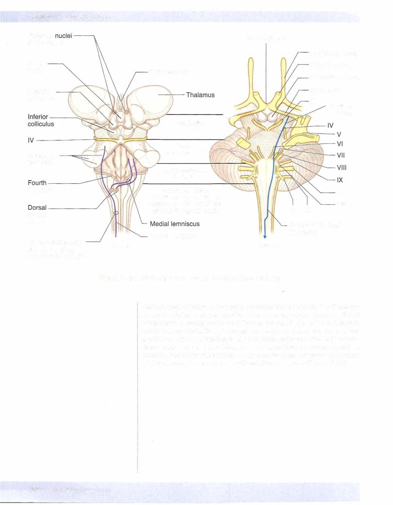
•This pathway conducts sensory information for touch, proprioception, vibration, and pressure. The primary afferent neurons ofthis pathway have their cell bodies in the dorsal root ganglia. Their axons enter the spinal cord and ascend in the dorsal columns of the white matter as the fasciculus gracilis (from lower limb) or the fasciculus cuneatus (from upper limb). They synapse with the second neuron in the same named nuclei in the lower medulla. Axons ofthe second neuron decussate (internal arcuate fibers) and ascend the midline of the brain stem in the medial lemniscus to reach the ventral posterolateral (VPL) nucleus of the thalamus. The third neuron then projects through the posterior limb ofthe internal capsule to the somatosensory cortex. Lesions above the decussation (in the brain stem or cortex) produce contralateral loss of joint position, vibration, and touch, whereas lesions below decussations (in the spinal cord) produce ipsilateral deficits below the level of the lesion. A positive Romberg test indicates lesions of the sensory input to the cerebellum.
Anterolateral System
•The anterolateral pathway carries pain and temperature sensations. The first neuron fibers enter the spinal cord and synapse in the dorsal horn with the second neurons. The first neuron often ascends or descends one or 2 segments before they synapse. The second neuron axons then
decussate (ventral white commissure) and ascend the spinal cord and form the spinothalamic tract in the lateral funiculus of the white matter. The spinothalamic tract ascends the lateral aspect ofthe brain stem and synapses in the VPL nucleus ofthe thalamus where the third neuron projects to the cortex. All lesions ofthe spinothalamic tract in the spinal cord, brain stem, or cortex produce contralateral loss of pain and temperature below the lesion. Note that a central cord lesion at the spinal canal (syringomyelia) produces bilateral loss of pain and temperature at the level ofthe lesion.
•Lesions ofthe spinal cord that involve the above-mentioned tracts include poliomyelitis, tabes dorsalis, amyotrophic lateral sclerosis, anterior spinal artery occlusion, subacute combined degeneration, syringomyelia, and Brown-Sequard syndrome.