Материал: Kaplan USMLE-1 (2013) - Anatomy

Внимание! Если размещение файла нарушает Ваши авторские права, то обязательно сообщите нам

Section IV • Neuroscience

SENSORYAND MOTOR NEURAL SYSTEMS

Each of the following 5 ascending or descending neural tracts, fibers, or fasciculi courses through the brain stem and will be found at every transverse sectional level.

Medial Lemniscus

The medial lemniscus (ML) contains the axons from cellbodies found in the dor­ sal column nuclei (gracilis and cuneatus) in the caudal medulla and represents the second neuron in the pathway to the thalamus and cortex for discriminative touch, vibration, pressure, and conscious proprioception. The axons in the ML cross the midline ofthe medulla immediately after emerging from the dorsal col­ umn nuclei. Lesions in the ML, in any part of the brain stem, result in a loss of discriminative touch, vibration, pressure, and conscious proprioception from the contralateral side of the body.

Spinothalamic Tract (Part ofAnterolateral System)

The spinothalamic tract has its cells of origin in the spinal cord and represents the crossed axons ofthe second neuron in the pathway conveying pain and tempera­ ture to the thalamus and cortex. Lesions of the spinothalamic tract, in any part of the brain stem, results in a loss of pain and temperature sensations from the contralateral side of the body.

CorticospinalTract

The corticospinal tract controls the activity oflower motoneurons, and interneu­ ron pools for lower motoneurons course through the brain stem on their way to the spinal cord. Lesions of this tract produce a spastic paresis in skeletal muscles of the body contralateral to the lesion site in the brain stem.

Descending Hypothalamic Fibers

The descending hypothalamic fibers arise in the hypothalamus and course with­ out crossing through the brain stem to terminate on preganglionic sympathetic neurons in the spinal cord. Lesions ofthis pathway produce an ipsilateral Horner syndrome. Horner syndrome consists of miosis (pupillary constriction), ptosis (drooping eyelid), and anhidrosis (lack of sweating) in the face ipsilateral to the side of the lesion.

Descending hypothalamic fibers course with the spinothalamic fibers in the lat­ eral part of the brain stem. Therefore, brain stem lesions producing Horner syn­ drome may also result in a contralateral loss of pain and temperature sensations from the limbs and body.

Medial Longitudinal Fasciculus

The medial longitudinal fasciculus is a fiber bundle interconnecting centers for hor­ izontal gaze, the vestibular nuclei, and the nerve nuclei of CN III, rv,and VI, which

innervate skeletal muscles that move the eyeball. This fiber bundle courses close to the dorsal midline ofthe brain stem and also contains vestibulospinalfibers, which

course through the medulla to the spinal cord. Lesions of the fasciculus produce internuclear ophthalmoplegia and disrupt the vestibulo-ocular reflex.
390 MEDICAL

MEDULLA

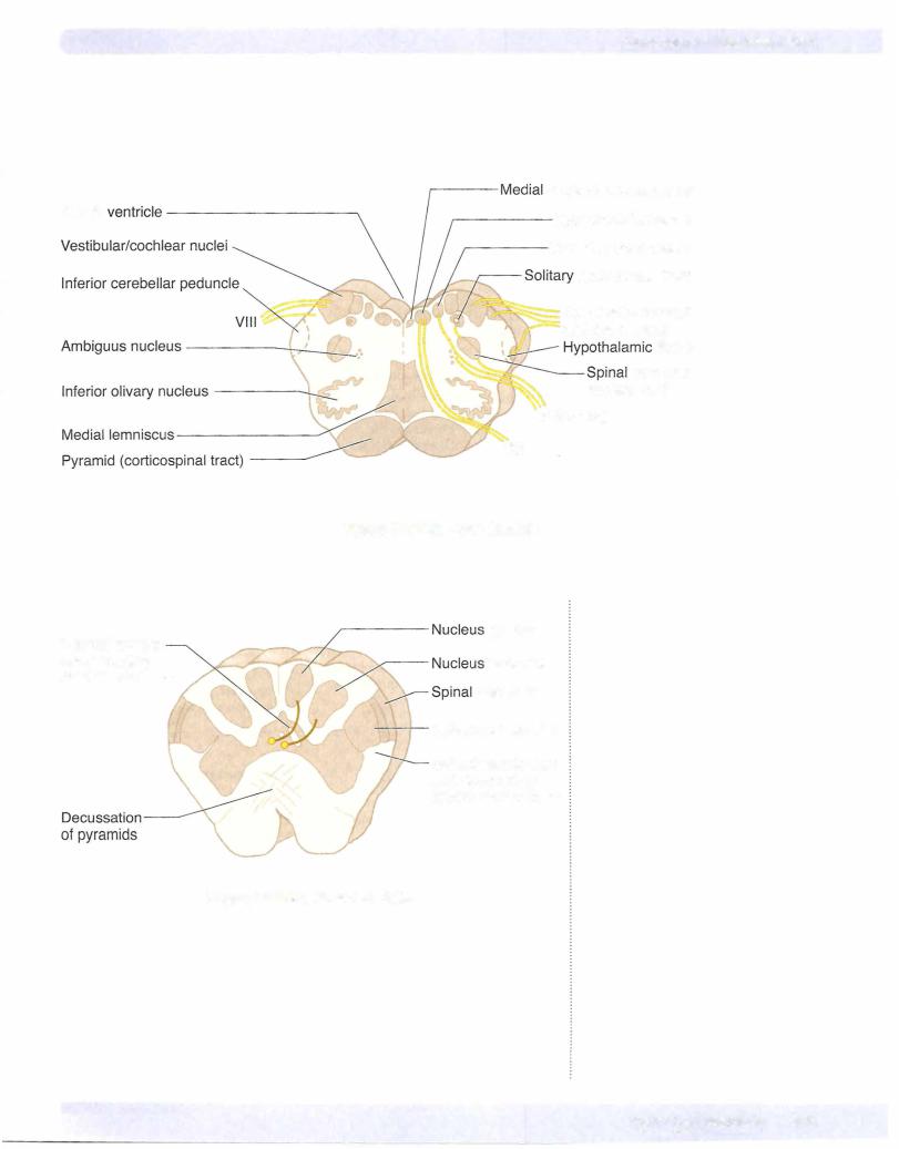
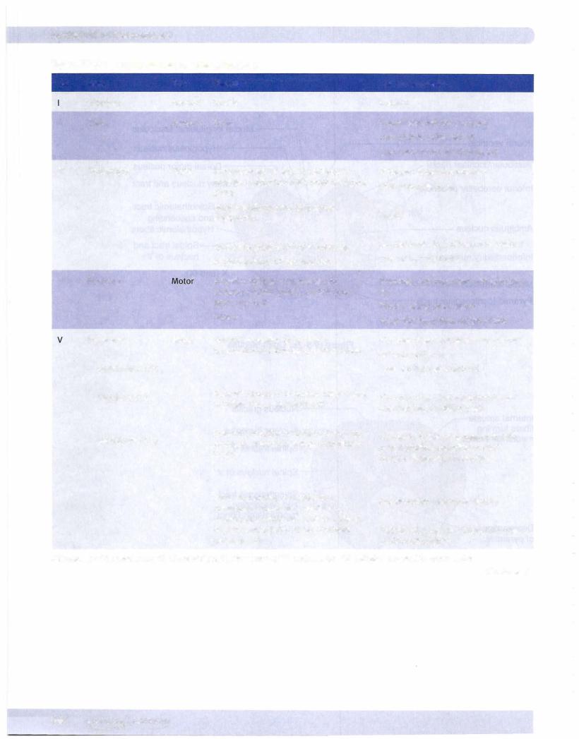
In the caudal medulla, 2 of the neural systems-the corticospinal and dorsal column-medial lemniscal pathways-send axons across the midline. The nucleus gracilis and nucleus cuneatus give rise to axons that decussate in the caudal me­ dulla (the crossing axons are the internal arcuate fibers), which then form and ascend in the medial lemniscus.

The corticospinal (pyramidal) tracts, which are contained in the pyramids, course ventromedially through the medulla. Most of these fibers decussate in the caudal medulla just below the crossing of axons from the dorsal column nuclei, and then travel down the spinal cord as the (lateral) corticospinal tract.

The olives are located lateral to the pyramids in the rostral two-thirds ofthe medul­ la. The olives contain the convoluted inferior olivary nuclei. The olivary nuclei send climbing (olivocerebellar) fibers into the cerebellum through the inferior cerebellar peduncle. The olives are a key distinguishing feature ofthe medulla.

The spinothalamic tract and the descending hypothalamic fibers course together in the lateral part of the medulla below the inferior cerebellar peduncle and near the spinal nucleus and tract of CN V.

Cranial Nerve Nuclei

Spinal nucleus ofV

The spinal nucleus of the trigeminal nerve (CN V) is located in a position analo­ gous to the dorsal horn of the spinal cord. The spinal tract ofthe trigeminal nerve lies just lateral to this nucleus and extends from the upper cervical cord (C2) to the point of entry of the fifth cranial nerve in the pons. Central processes from cells in the trigeminal ganglion conveying pain and temperature sensations from the face enter the brain stem in the rostral pons but descend in the spinal tract of CN V and synapse on cells in the spinal nucleus (Figure IV-5-3).

Solitary nucleus

The solitary nucleus receives the axons of all general and special visceral afferent fibers carried into the CNS by CN VII, IX, and X. These include taste, cardiore­ spiratory, and gastrointestinal sensations carried by these cranial nerves. Taste and visceral sensory neurons allhave their cell bodies in ganglia associated with CN VII, IX, and X outside the CNS.

Nucleus ambiguus

The nucleus ambiguus is a column of large motoneurons situated dorsal to the inferior olive. Axons arising from cells in this nucleus course in the ninth and tenth cranial nerves. The component to the ninth nerve is insignificant. In the tenth nerve, these fibers supply muscles of the soft palate, larynx, pharynx, and upper esophagus. A unilateral lesion will produce ipsilateral paralysis of the soft palate causing the uvula to deviate away from the lesioned nerve and nasal regur­ gitation of liquids, weakness of laryngeal muscles causing hoarseness, and pha­ ryngeal weakness resulting in difficulty in swallowing.

Chapter 5 • The Brain Stem

MEDICAL 391

Источник: https://studfile.net/preview/14638320/