Материал: Kaplan USMLE-1 (2013) - Anatomy

Внимание! Если размещение файла нарушает Ваши авторские права, то обязательно сообщите нам

Section IV • Neuroscience

Dorsal motornucleus ofCNX

These visceral motoneurons ofCN X arelocated lateral to the hypoglossal nucleus in the floor ofthe fourth ventricle. This is a major parasympathetic nucleus ofthe brain stem, and it supplies preganglionic fibers innervatingterminalganglia in the thorax and the foregut and midgut parts ofthe gastrointestinal tract.

Hypoglossal nucleus

The hypoglossal nucleus is situated near the midline just beneath the central ca­ nal and fourth ventricle. This nucleus sends axons into the hypoglossal nerve to innervate all ofthe tongue muscles except the palatoglossus.

The accessory nucleus

The accessorynucleus is found in the cervical spinal cord. The axons ofthe spinal accessory nerve arise from the accessory nucleus, pass through the foramen mag­ num to enter the cranial cavity, and join the fibers ofthe vagus to exit the cranial cavity through the jugular foramen. As a result, intramedullary lesions do not affect fibers ofthe spinal accessory nerve. The spinal accessory nerve supplies the sternocleidomastoid and trapezius muscles.

Therootlets ofthe glossopharyngeal (CN IX) andvagus (CN X) nerves exitbetween the olive and the fibers of the inferior cerebellar peduncle. The hypoglossal nerve (CN XII) exits more mediallybetween the olive and the medullarypyramid.

PONS

The pons is located between the medulla (caudally) and the midbrain (rostrally). The cerebellum overlies the pons. It is connected to the brain stem by 3 pairs of cerebellar peduncles. The fourth ventricle is found between the dorsal surface ofthe pons and the cerebellum. The ventral surface ofthe pons is dominated by fibers, which form a large ventral enlargement that carries fibers from pontine nuclei to the cerebellum in the middle cerebellar peduncle. This ventral enlarge­ ment is the key distinguishing feature ofthe pons.

The corticospinal tracts are more diffuse in the pons than in the medulla and are embedded in the transversely coursing fibers that enter the cerebellum in the middle cerebellar peduncle.

The medial lemniscus is still situated near the midline but is now separated from the corticospinal tracts by the fibers forming the middle cerebellar peduncle. The mediallemniscus has changed from a dorsoventral orientation in the medulla to a more horizontal orientation in the pons.

The spinothalamic tract and the descending hypothalamic fibers continue to course together in the lateral pons.

The lateral lemniscus, an ascending auditory pathway, is lateral and just dorsal to the medial lemniscus. The lateral lemniscus carries thebulkofascending auditory fibers from both cochlear nuclei to the inferior colliculus ofthe midbrain.

The medial longitudinal fasciculus (MLF) is located near the midlinejustbeneath the fourth ventricle.

392 M E D I CAL

Chapter 5 • The Brain Stem

Cranial Nerve Nuclei

Abducens nucleus

The abducens nucleus is found near the midline in the floor of the fourth ven­ tricle just lateral to the MLF.

Facial motornucleus

The facial motor nucleus is located ventrolateral to the abducens nucleus. Fibers from the facial nucleus curve around the posterior side of the abducens nucleus (the curve forms the internal genu of the facial nerve), then pass ventrolaterally to exit the brain stem at the pontomedullary junction.

Superiorolivary nucleus

The superior olivary nucleus lies immediately ventral to the nucleus of CN VII and receives auditory impulses from both ears by way ofthe cochlear nuclei. The cochlear nuclei are found at the pontomedullary junction just lateral to the infe­ rior cerebellar peduncle.

Vestibular nuclei

The vestibular nuclei are located near the posterior surface of the pons lateral to the abducens nucleus, and extend into the medulla.

Cochlearnuclei

The dorsal and ventral cochlear nuclei are found at the pontomedullary junction.

All of the fibers of the cochlear part of CN VIII terminate here.

Trigeminal nuclei

Motor Nucleus-Pons

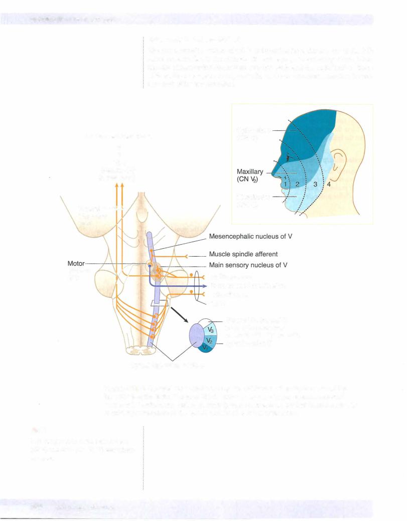
The motor nucleus of CN V is located in the pons just medial to the main sensory nucleus ofthe trigeminal and adjacent to the point ofexit or entry ofthe trigeminal nerve fibers. These motor fibers supply the muscles ofmastication (masseter, tem­ poralis, and medial and lateral pterygoid (Figure IV-5-3).

Main Sensory Nucleus-Pons

The main sensory nucleus is located just lateral to the motor nucleus.

The main sensory nucleus receives tactile and pressure sensations from the face, scalp, oral cavity, nasal cavity, and dura.

Spinal Trigeminal Nucleus-Spinal cord to pons

The spinal trigeminal nucleus is a caudal continuation ofthe main sensory nu­ cleus, extending from the mid pons through the medulla to the cervical cord. Central processes from cells in the trigeminal ganglion conveying pain and tem­ perature sensations from the face descend in the spinal tract ofV and synapse on cells in the spinal nucleus.

MEDICAL 393

Chapter 5 • The Brain Stem

Cranial NervesV, VI, VII, and VIII

Four cranial nerves emerge from the pons. Cranial nerves VI, VII, and VIII emerge from the pontomedullary junction. The facial nerve is located medial to the vestibu­ locochlear nerve. The abducens nerve (CN VI) emerges near the midline lateral to the corticospinal tract. The trigeminal nerve (C V) emerges from the middle of the pons.

MIDBRAIN

The midbrain (mesencephalon) is located between the pons and diencephalon. The cerebral aqueduct, a narrow channel that connects the third and fourth ven­ tricles, passes through the midbrain. The inferior colliculi and superior colliculi are found on the dorsal aspect of the midbrain above the cerebral aqueduct. The inferior colliculus processes auditory information received bilaterally from the co­ chlear nuclei by axon fibers of the lateral lemniscus. The superior colliculi help direct movements ofboth eyes in gaze. The pretectal region is locatedjust beneath the superior colliculi and in front of the oculomotor complex. This area contains interneurons involved in the pupillary light reflex. The massive cerebral peduncles extend ventrally from the midbrain. 1be cerebral peduncles contain corticospinal and corticobulbar fibers. The interpeduncular fossa is the space between the cere­ bral peduncles.

The substantia nigra is the largest nucleus of the midbrain. It appears black to dark brown in the freshly cut brain because nigral cells contain melanin pig­ ments. Neurons in the substantia nigra utilize Dopamine and GABA as neu­ rotransmitters.

The medial lemniscus and spinothalamic tract and descending hypothalamic fi­ bers course together ventrolateral to the periaqueductal gray.

The MLF continues to be located near the midline, just beneath the cerebral aq­ ueduct.

The mesencephalic nuclei ofthe trigeminal nerve are located on either side ofthe central gray.

Cranial Nerve Nuclei

The trochlear nucleus is located just beneath the periaqueductal gray near the midline between the superior and inferior colliculi. The oculomotor nucleus and the nucleus of Edinger-Westphal are found just beneath the periaqueductal gray near the midline at the level of the superior colliculi.

Two cranial nerves emerge from the midbrain: the oculomotor (CN III) and the trochlear (C IV) nerves.

The oculomotor nerve arises from the oculomotor nucleus and exits ventrally from the midbrain in the interpeduncular fossa. CN III also contains preganglion­ ic parasympathetic axons that arise from the nucleus of Edinger-Westphal, which lies adjacent to the oculomotor nucleus.

Axons of the trochlear nerve decussate in the superior medullary velum and exit the brain stem near the posterior midline just inferior to the inferior colliculi.

MEDICAL 395

Источник: https://studfile.net/preview/14638320/