Материал: Kaplan USMLE-1 (2013) - Anatomy
Connective Tissue |
2 |
Connective tissue (CT) provides supportforparenchymalcells (epithelial andother functional cell types) that carry out the specific tissue and organ functions. Paren chymal cells are not only attached to and supported by connective tissue, they also have active mechanical, chemical, and other interactions between parenchymal cells and components of the connective tissue. CT is comprised of support cells, guest cells, and their associated extracellularmatrix (ECM) or ground substance.
CopyrightMcGraw-Hi// Companies. Usedwithpermission.
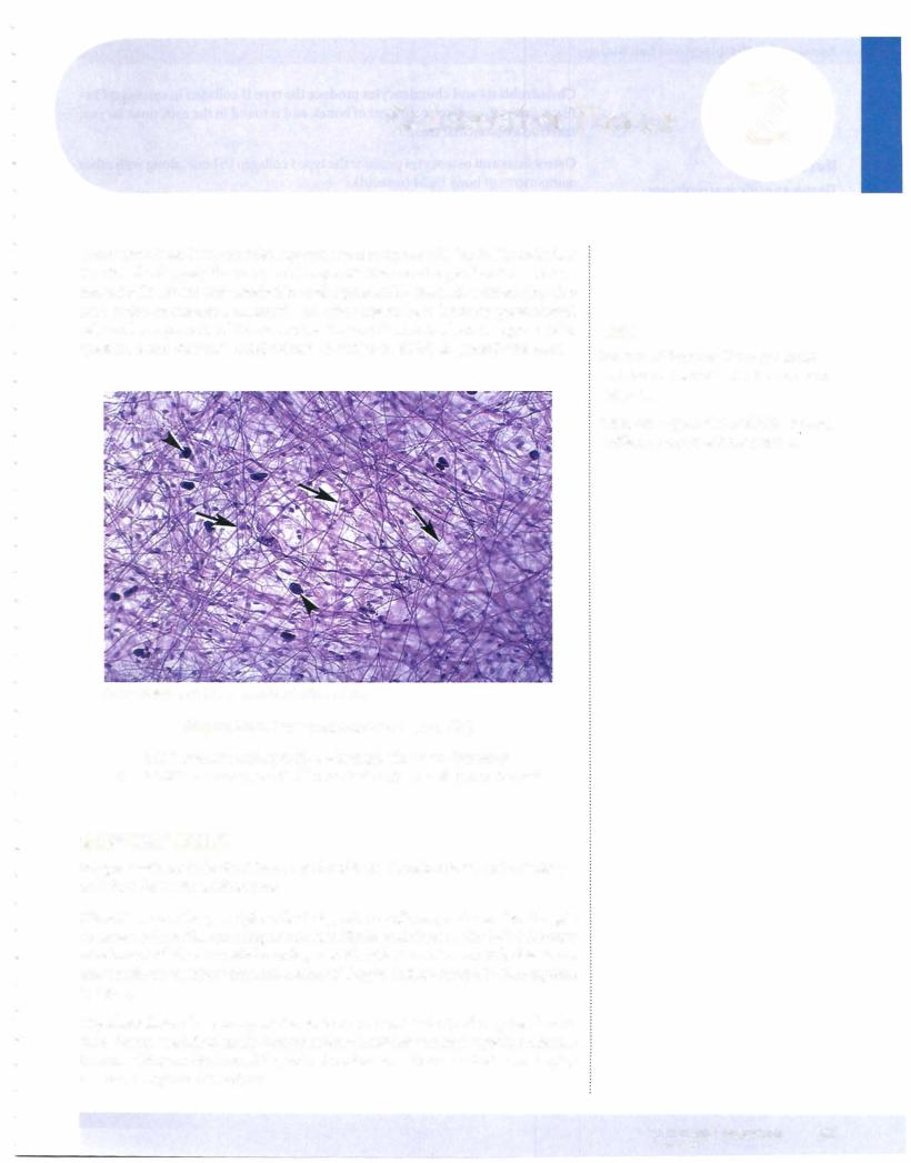
Figure 1-2-1. Loose connective tissue (LCT)
LCT contains collagen fibers and elastic fibers (arrows); mast cells are one type of CT permanent guest cell (arrowheads)
SUPPORT CELLS
Support cells include fibroblasts, myofibroblasts, chondroblasts, and osteoblasts and their derivatives, adipocytes.
Fibroblasts are the principle cells that produce collagen, and are also thought to have a role in the ordering of collagen fibrils and fibers in the ECM through attachment ofthe extracellular collagen to fibroblast membranes. Fibroblasts are more resistant to injury than most other cell types and are crucial in the response to injury.
Myofibroblasts share many of the features of fibroblasts (and may be derived from them), but additionally contain actin microfilaments and myosin thick fila ments, adding an intrinsic ability ofcell contraction. This is critical in reshaping tissue as it repairs after injury.
Note
There are 2 types of CT proper: loose and dense. Dense CT may be regular or irregular.
There are 4 types of special CT: adipose, cartilage, bone, and bone marrow.
MEDICAL 27
Section I • Histology and Cell Biology
Note
Tissue-specific macrophages:
Kupffer cells-liver
Osteoclasts-bone
Microglia-CNS
Chondroblasts and chondrocytes produce the type II collagen in cartilage. Car tilage covers the articular surfaces ofbones, and is found in the ears, nose, larynx, and tracheobronchial tree.
Osteoblasts andosteocytes produce the type I collagen in bone, along with other components of bone ECM (osteoid).
Adipocytes are the principal support cell in fat (adipose tissue) and may be found in other organs as well. There are 2 basic types of fat cells. In the adult, all or most fat is white fat or unilocular fat. A single adipocyte contains a single huge vacuole of fat (not membrane-bound), which pushes and flattens the cell nucleus to one side. Adipose cells have a surrounding external lamina and type III reticulin collagen fibers are found between the adipocytes. Brown fat or mul tilocular fat is a second type of fat found in fetuses and neonates (occasionally in small amounts in adults) that is composed of adipocytes with multiple small vacuoles offat and with a round central nucleus. This type of fat is thought to be important in generating heat in the newborn.
PERMANENT GUEST CELLS
The permanentguestcells are macrophages and mast cells.
Macrophages (histiocytes) are phagocytic cells derived from bone marrow monocytes, which can leave the bloodstream, enter tissues, and remain there for long periods. Macrophages are motile, (they can move within the tissue), are phagocytic, and are a source of metalloproteinases, which can break down ECM components They can process and present antigens to cells of the immune sys tem, either locally or by travelling to distant sites of immune cell accumulation. Tissue macrophages can resemble fibroblasts.
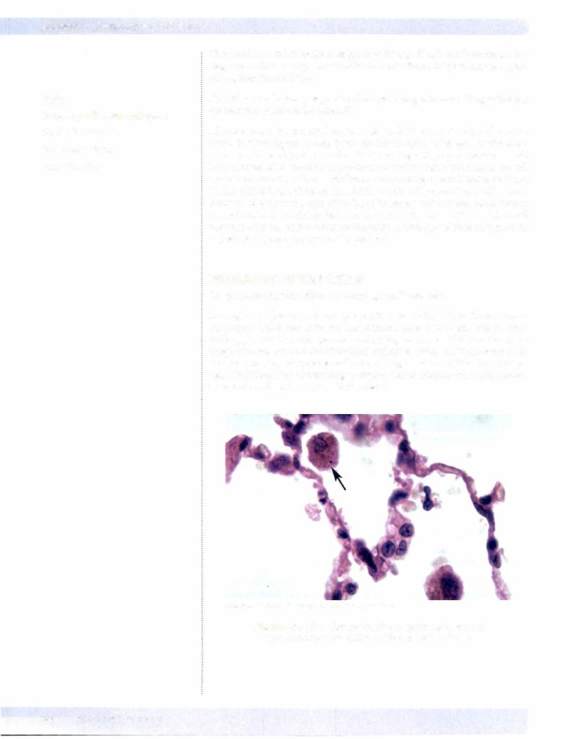
Copyright McGraw-Hill Companies. Used with permission.
Figure 1-2-2. Alveolar macrophage (arrow) is a second type of permanent guest cell in connective tissue
28 MEDICAL
Chapter 2 • Connective Tissue
Mast cells are closely related but not derived from blood basophils, and store inflammatorymediators such as histamine that have an early role in response to tissue injury, particularly immune mediated injury associated with immediate hypersensitivity reactions. IgE bound to the surface membrane ofthe mast cells mediates these reactions. Morphologically, mast cells contain large, membrane bound granules of their inflammatory mediators, which can best be visualized with special stains in the light microscope.
TRANSIENT CELLS
Transient cells migrate into connective tissue from the blood and include lym phocytes, plasma cells, neutrophils, eosinophils, basophils, and monocytes.
EXTRACELLULAR MATRIX
The extracellular matrix consists offibrillar proteins (collagen, elastin, fibrillin, fibronectin), proteoglycans that are associated with water and form a gel matrix, and glycoproteins that form structural proteins and include laminin, entactin, and tenascin.
Fibrillar proteins are derived from soluble precursors synthesized and secreted by support cells ofthe CT. The most abundantis collagen, butvarious connective tissues may also have a variable content of elastin and fibrillin, and fibronectin, which help to anchor cells to basal lamina.
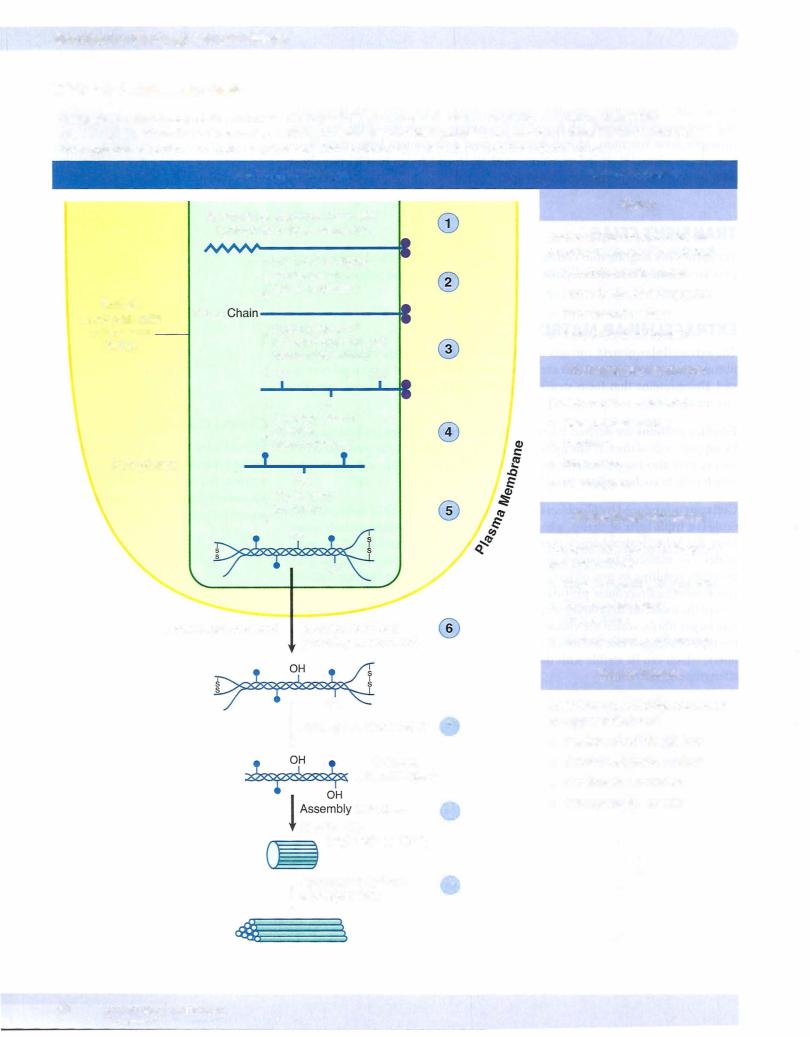
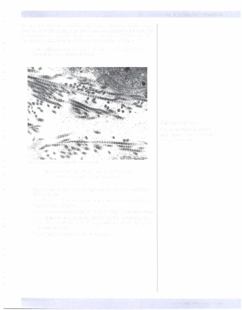
Collagen is produced by fibroblasts and myofibroblasts. The initial collagen mol ecule is a triple helix ofchains, which are tightly bound and which exclude water from the helix (hydrophobic), thereby allowing very tight packing of the mol ecules. The molecule undergoes glycosylation and hydroxylation ofvarious ami no acids, resulting in procollagen. Procollagen is secreted into the extracellular space, where extracellular peptidases (also offibroblast origin) cleave off a por tion ofthe molecule toproducetropocollagen. Tropocollagen then selfassembles into larger fibrils, which are visible with the electron microscope. Depending on the type ofcollagen and the tissue, fibrils may assemble into even larger collagen fibers, which can be visible with the light microscope. Collagen provides tensile strength to tissue.
Clinical Correlate
lgE-sensitized mast cells degranulate during hypersensitivity reactions (asthma, hay fever, eczema). The release of histamine during asthma causes difficulty breathing.
MEDICAL 29
There are at least 25 different collagen chain types, and they can combine to pro duce at least 19 different collagens. Some form fibrils, others are associated with but are not the backbone of the fibrils, and yet others form a felt-like mesh rather than fibrils. An abbreviated list of the most important ones includes:
•Type I collagen is found in skin, tendon, ligaments, bone, and cornea. Most of the body's collagen is Type I.
Copyright McGraw-Hill Companies. Used with permission.
Figure 1-2-3. Collagen fibers with a banding pattern with repeating units of 64 nm in length.
•Type II collagen is found in cartilage, intervertebral discs, and vitreous body of the eye.
•Type III collagen is found in blood vessels, and forms reticular fibers in various organs and tissues.
-Links the basal lamina of cells to the underlyingECM in many tissues.
-Comprises the main supporting fibers in the ECM surrounding the
parenchymal cells of lymphoid (lymph node and spleen) organs, bone marrow, and liver
• Type IV collagen is found in the basal lamina.
Chapter 2 • Connective Tissue
Clinical Correlate
Type Ill is the first collagen type synthesized in wound healing, and is then replaced by type I.
MEDICAL 31