Материал: Kaplan USMLE-1 (2013) - Anatomy
Chapter 3 • Cartilage and Bone
BONE
Bone is a unique connective tissue in that it not only has cells and ECM called osteoid including type I collagen and GAGs, but also the matrix is calcified and rigid. This requires adaptations ofthe resident cells not seen in most tissues. Unlike cartilage, bone is vascularized.
Bone Cells
Osteoblasts secrete bone ECM (osteoid). Osteoblasts are derived from osteo progenitor cells, which are formed from multipotent mesenchymal stem cells, the same cells that form allconnective tissue. These osteoprogenitor cells may be carried into preexisting cartilage in the connective tissue from surrounding blood vessels that grow into cartilage during embryogenesis or from connective tissue residing in the bone marrow spaces during subsequent bone growth and remodeling.
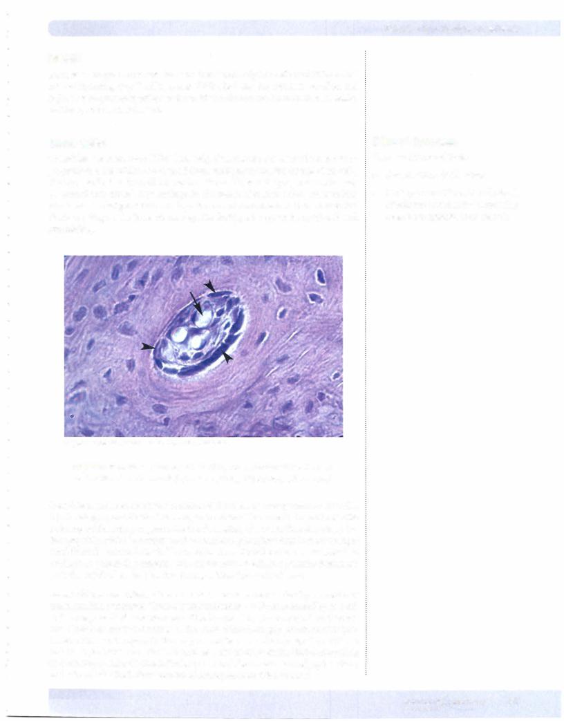
Copyright McGraw-Hill Companies. Usedwith permission.
Figure 1-3-3. Haversian canal lined by osteoprogenitor cells and osteoblasts (arrowheads) and containing blood vessels (arrow)
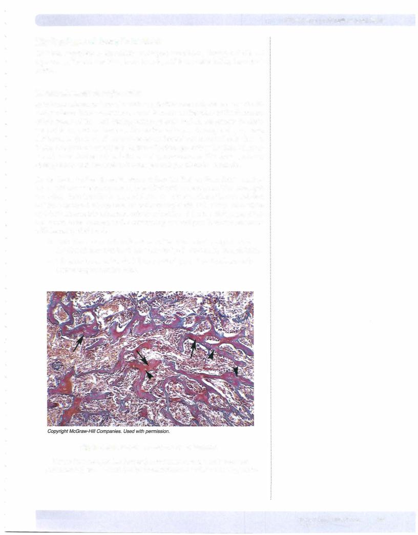
Osteoblasts are specialized to synthesize and secrete the components ofosteoid, type I collagen, and GAGs. The unique structure ofthe osteoid, in particular the ordering of the collagen, promotes the formation of a crystalline structure, hy droxyapatite, which is comprised ofcalcium and phosphate ions that are precipi tated from the extracellular fluid. Osteoblasts usually adhere to a surface, such as cartilage or preexisting osteoid, and secrete new osteoid in a polarized manner onto the attached surface, leading to ofbone.
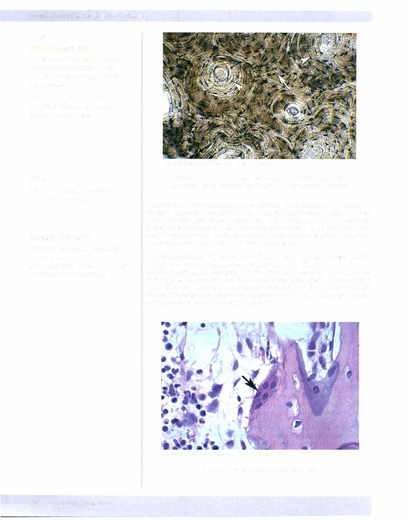
As osteoblasts are added, other osteoblasts become surrounded by osteoid and transform into osteocytes. These osteoblasts become totally surrounded byosteoid, and areresponsible formaintenanceofthe bonematrix. Osteocytesextendthrough canaliculi that are tiny channels in the osteoid that form gap junctions with pro cesses ofother osteocytes. In this way, osteocytes can exchange signals, nutrients, and wasteproducts. There is also a small amount ofextracellular fluid surrounding each osteocyte and its intracanalicularprocesses. The combination ofgapjunctions and extracellular fluid allow survival ofosteocytes embedded in bone.
Clinical Correlate
There are 2 forms of bone:
•Compact bone (solid mass)
•Spongy or cancellous bone (network of spicules or trabeculae separating spaces occupied by bone marrow)
MEDICAL 37
Chapter 3 • Cartilage and Bone
Mechanisms of Bone Formation
All bone, regardless of formation, undergoes remodeling throughout life. All
3 processes, bone formation, bone growth, and bone remodeling, have simi larities.
lntramembranous boneformation
In intramembranous bone formation, primitive mesenchyme can give rise di rectly to bone. Intramembranous bone formation takes place in the formation of flat bones of the skull during embryogenesis, and in the growth in thick ness of dense cortical bone on the surface of bones. During embryogenesis, flat bones begin as small collections of condensed mesenchymal cells that are induced to become osteoblasts. As the collections grow they fuse into intercon nected cords (trabecula) with intervening mesenchyme. This forms primary
and has randomly arranged collagen fibers in its matrix.
As the bone further develops, some trabeculae fuse to form dense cortical bone, without intervening large spaces filled with mesenchyme. The cortex gets remodeled into lamellar bone, and forms the outer surface of bones and does not have cavities for bone marrow and hematopoiesis. Other trabeculae widen and form an anastomosing network of trabeculae with intervening spaces that can house bone marrow, as the intervening mesenchyme becomes populated with hematopoietic cells.
•This is known as trabecular or cancellous bone, and comprises the interior ofmost bones, whose outer surface is covered by cortical bone.
•Thus, flat bones ofthe skull have 2 outer layers of cortical bone with intervening trabecular bone.
Figure 1-3-6. lntramembranous bone formation
Newly formed spicules (arrows) containing osteocytes in lacunae (arrowheads) are surrounded by mesenchyme of primary spongy bone
MEDICAL 39

Section I • Histology and Cell Biology
Clinical Correlate
Rickets results from calcium deficiency during bone growth and may be due to insufficient dietary Ca+ or vitamin D.
Endochondral boneformation
In endochondralbone formation, bone is formed on the template ofpreexisting hyaline cartilage. Endochondral bone formation occurs in long bones ofthe ex tremities and in vertebrae and bones ofthe pelvis, and starts as ahyaline cartilagi nous template that is formed from chondroblast and chondrocyte differentiation from chondroid progenitor cells derived from mesenchyme.
the surface of preexisting cartilage is endo
• In long bones, this initially takes place in the center of the shaft (diaphy sis) of the cartilage template and is the primary center ofossification.
This leads to the progressive replacement of the inner cartilage with trabecular bone, which progressively is laid down toward the ends of the bones, leading to increase in bone length.
•At the same time, other osteoprogenitor cells at the surface periosteum begin to lay down osteoid as a layer on the outer surface ofthe cartilage template, and this form of intramembranous bone formation will ulti mately form the outer cortex of the long bones.
•Later in development, secondary (or late) centers ofossification form separately through a process of cartilage hypertrophy, ingrowth ofves sels from outside the cartilage template, and replacement of cartilage by new trabecular bone and marrow. This takes place at the ends (epiphy ses) of long bones.
•In long bones, most but not all ofthe cartilage is replaced by bone and marrow.
•At the ends of the bones that will form synovial joints, the cartilage per sists; and at the interface of the epiphysis with the diaphysis, some ofthe cartilage persists as the epiphyseal growth plate, important in the elon gation of bones.
•Depending on the complexity of shape ofbones, there is some variation
in the formation of primary and secondary centers ofossification. Both the initial surface dense corticalbone and the trabecular bone in the ossi fication centers is woven bone, later to be remodeled into lamellar bone.
In long bones, a band of cartilage is present at each end of the diaphysis and persists even after appearance of the secondary center of ossification and until the bone reaches its ultimate length. The progressive growth of this band of cartilage toward the ends of the bone by a combination of chondrocyte prolif eration and interstitial growth of chondroid matrix is the mechanism bywhich the bone lengthens.
•Advancing cartilage is replaced by new bone, which advances behind the growing epiphyseal plate.
•At puberty, the bone growth eventually catches up and completely replaces this cartilage, halting the growth and closing the epiphyseal plate, an event recognizable on x-ray by loss of the previous relatively lucent band of cartilage near the epiphyseal-diaphyseal interface.
40 MEDICAL
Chapter 3 • Cartilage and Bone
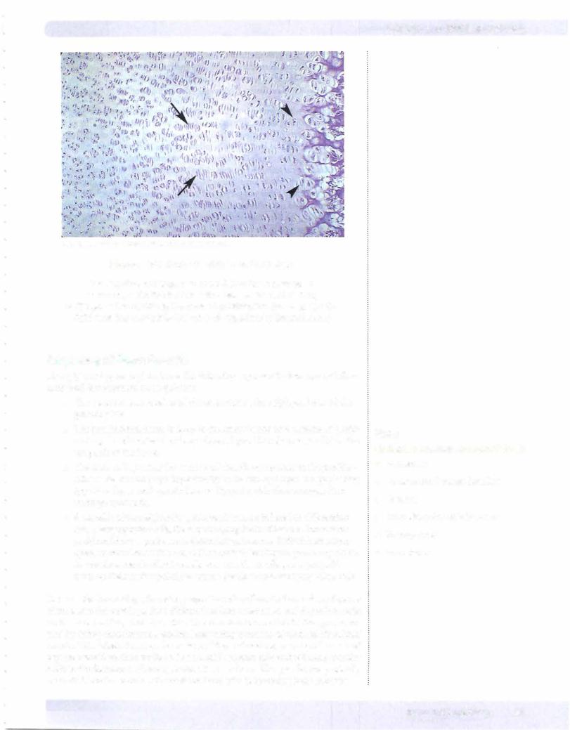
CopyrightMcGraw-Hill Companies. Used with permission.
Figure 1-3-7. Endochondral Bone Formation
The hyaline cartilage is to the left and bone marrow is to the right. On the far left is the reserve zone of resting
cartilage. In the middle is the zone of proliferation (arrows). On the right near the marrow is the zone of hypertrophy (arrowheads).
Sequence of Bone Growth
The epiphyseal growth plate shows the following sequence for bone growth from early fetal development up to puberty.
•The reserve zone consists of chondrocytes at the epiphyseal end of the growth plate.
•The proliferatingzone is deep to the reserve zone and consists of prolif erating chondrocytes that form clones aligned in columns parallel to the long axis of the bone.
•The zone ofhypertrophy consists of chondrocytes deep to the prolifer ating zone that undergo hypertrophy by becoming larger and producing type X collagen and growth factors. Hypertrophic chondrocytes then undergo apoptosis.
•Stem cells of the neighboring perichondrium are induced to differentiate into osteoprogenitor cells, thereby changing the limiting membrane from perichondrium to periosteum. Osteoclast precursors, derived from mono cytes, are recruited to this site,with osteoclasts leading the process by attack ing cartilage matrix. Blood vessels grow into the cartilage, carrying with them on their surface primitive connective tissue and osteoprogenitor cells.
Once inside the cartilage, the osteoprogenitor cells adhere to the surface ofspaces cleared into the cartilage, then differentiate into osteoblasts and deposit osteoid on the new cartilage matrix surface. The invasion occurs first in the spaces cre ated by dying chondrocytes, leaving intervening columns of calcified chondroid matrix. Osteoblasts form on the surface of these chondroid matrix columns and deposit osteoid on their surfaces. The osteoid becomes mineralized and gradually leads to the formation ofnew spicules ofwoven bone. The spicules are gradually converted into trabeculae of cancellous bone with intervening bone marrow.
Note
Cortical or lamellar bone consists of:
•Periosteum
•Outer circumferential lamellae
•Osteons
•Inner circumferential lamellae
•Spongy bone
•Endosteum
MEDICAL 41