Материал: Kaplan USMLE-1 (2013) - Anatomy
Section I • Histology and Cell Biology
Clinical Correlate
In Marfan syndrome, elastic tissue is weakened.
•Ocular defects include myopia and detached lens.
•Skeletal defects are long arms, legs, fingers, and scoliosis.
•Cardiovascular defects are mitral valve prolapse and dilated aorta.
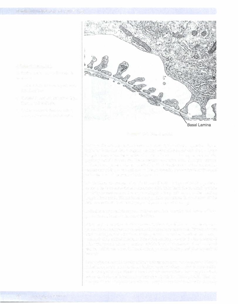
Figure 1-2-4. Basal lamina
Collagen fibers in connective tissue can have a highly variable disposition, form ing loose irregular, dense regular, or dense irregular connective tissue. Loose irregular connective tissue is found in the submucosa of many organs and the papillary dermis of the skin. Dense regular connective tissue is highly ordered and may be organized in larger parallel bundles in tendons or in other ordered ar rangements such as bone and cornea. Dense irregular connective tissue is found in the dermis and capsules of various organs.
Elastic fibers are composed of elastin and fibrillin, 2 types of fibrillar proteins secreted by connective tissue supporting cells,. They form fibers, which provide elasticity or resilience to tissues, meaning that they will return to their resting length after stretch. Elastic fibers are especially prominent in the dermis of the skin, the walls of blood vessels, especially arteries, and the lung.
Reticular fibers contain type III collagen and form capsules and stroma oflym phatic tissues, bone marrow, and the liver.
Fibronectin is another fibrillar protein in the extracellular matrix, again com prised ofinsoluble monomers secreted by connective tissue cells. Fibronectin can bind to collagen and other matrix components, as well as to cell-surface recep tors, including epithelial cells, and therefore are important in the interactions of cells with matrix. There is another soluble form of fibronectin found in blood plasma, which has a role in blood clotting, wound healing and phagocytosis of bacteria.
Proteoglycans consist mostly of long unbranched glycosaminoglycans (GAGs) linked to a protein core. GAGs are highly negatively charged, due to many sulfat ed residues, attract positively charged cations around them. Proteoglycans form aggregates by being linked to a hyaluronan molecule by a linker protein. There are 5 major classes of glycosaminoglycans. They include hyaluronic acid (the only
32 M EDICAL
one that does not form proteoglycans), chondroitin sulfate, dermatan sulfate, keratan sulfate and heparan sulfate. The major components ofECM include a gel formed by the interaction ofwater with proteoglycan aggregates. This gel resists compressive forces and provides a medium through which water soluble com pounds can diffuse such as between blood vessels and parenchymal cells, includ ing ions, nutrients, metabolites, growth factors, hormones, etc.
Glycoproteins have a relatively greater content of protein compared to proteo glycans. Laminin, entactin, and tenascin are non-filamentous structural glyco proteins in the ECM.
•Laminin is a component ofthe basement membrane of most epithelial and endothelial cells.
•Laminin binds integrins of the cell membrane and link the cell mem brane to the basal lamina via entactin, which links laminin to the type IV collagen of the basal lamina.
•Tenascin is another important adhesion molecule in the cell.
Ifthese 2 conditionsare notpresent,fibroblasts replacethe damaged parenchyma with scar tissue. Fibroblasts can be stimulated to proliferate and produce large amounts of extracellular collagen to fillin the defect. In either case, in regen eration or scar formation, macrophages clear away any debris of damaged cells and ECM components. Macrophages may already reside in the tissue or can be recruited from the blood stream. The recruitment, proliferation, and function of cells involved in wound healing is directed by the sequential expression and sup pression ofvarious growth factors and inflammatory mediators.
Chapter 2 • Connective Tissue
CUnical Correlate
If an organ is damaged, it may be able to restore itselfthrough regeneration totally or nearly to the state it was in prior to injury. Regeneration requires a resident population of parenchymal cells ableto replace lost ones, either stem cells or differentiated cells that still have the ability to proliferate.
Regeneration requires an intact underlying connective tissue support network, including an intact basal lamina, to guide the organization of newly formed parenchymal cells.
MEDICAL 33
Section I • Histology and Cell Biology
ChapterSummary
•Connective tissue (Cl) provides support for parenchymal cells (epithelial and other functional cell types) that carry out the specific tissue and organ functions.
•Connective tissue is comprised of support cells, guest cells, and their associated extracellular matrix (ECM) or ground substance. Support cells include fibroblasts, myofibroblasts, chondroblasts, osteoblasts and their derivatives, adipocytes. The permanent guest cells are macrophages and mast cells.
•The extracellular matrix consists offibrillar proteins (collagen, elastin, fibrillin, fibronectin), proteoglycans that are associated with water and form a gel matrix, and glycoproteins that form structural proteins and include laminin, entactin, and tenascin.
•Fibrillar proteins are mainly collagen, but various connective tissues may also have a variable content of elastin and fibrillin, and fibronectin, which help to anchor cells to basal lamina. Collagen is produced by fibroblasts and myofibroblasts.
•Type I collagen is found in skin, tendon, ligaments, bone, and cornea.
•Most of the body's collagen is type I. Type II collagen is found in cartilage,
intervertebral discs, and in vitreous body of the eye.
•Type Ill collagen is found in blood vessels, and it forms reticular fibers in various organs and tissues. It links the basal lamina ofcells to the underlying extracellular matrix in many tissues. Type Ill collagen comprises the main supporting fibers in the extracellular matrix surrounding the parenchymal cells of lymphoid (lymph node and spleen) organs, bone marrow, and liver.
•Type IV collagen is found in the basal lamina.
•Elastic fibers are composed of elastin and fibrillin and are especially prominent in the dermis ofthe skin, the walls of blood vessels, especially arteries, and the lun
•Fibronectin can bind to collagen and other matrix components, as well as to cell surface receptors, including epithelial cells, and therefore are important in the interactions of cells with matrix.
•Proteoglycans consist mostly of long unbranched glycosaminoglycans
(GAGs) linked to a protein core.
•Glycoproteins have a relatively greater content of protein compared to proteoglyc.ans. Laminin, entactin, and tenascin are non-filamentous structural glycoproteins in the ECM.
34 MEDICAL
Cartilage and Bone |
3 |
CARTILAGE
Cartilage, like all connective tissue, consists of cells and extracellular matrix (ECM). The ECM is rich in type II collagen fibrils and glycosaminoglycans (GAGs). Some specialized types of cartilage may have additional components in the matrix. Cartilage is avascular, and it can serve as the template for bone formation.
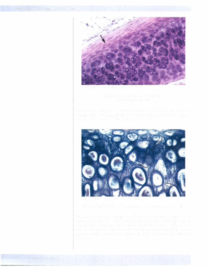
Cartilage formation takes place by differentiation ofmultipotential mesenchymal cells into chondroid precursor cells, which give rise to chondroblasts, which give rise to chondrocytes. The cartilage mass is surrounded by perichondrium, a membrane of connective tissue containing the stem cells which can give rise to both fibroblasts and chondroid progenitor cells. The chondroid progenitor cells change from small elongated cells to chondroblasts, which are more oval shaped, with more cytoplasm. They secrete type II collagen and some GAGs to produce extracellular chondroid matrix. The immature matrix initially surrounding chon droblasts is relatively acidophilic (pink staining), as it has proportionately more collagen compared to GAGs. As the chondroblast matures into a more rounded chondrocyte trapped in a lacuna within the matrix, the matrix also matures with proportionately more GAGs compared to collagen and more basophilic (blue staining). As the mass ofcartilage is formed, it is not penetrated by blood vessels.
Cartilage can enlarge in 2 ways. In appositionalgrowth, new cells can be added from the outer perichondrium. In internal or interstitial growth, the chondro cytes embedded deep within the cartilage can continue to produce additional ECM.
Chondrocytes are not polarized and can secrete new matrix circumferentially, in contrast to osteoblasts, which secrete only in one direction, adding onto a sur face. The interstitial growth depends more on continuing extracellular secretion and less on cell proliferation. Typically there are or 4 chondrocytes within a lacuna.
Types ofCartilage
There are 3 types of cartilage, all containing type II collagen and GAGs. Some times they have additional extracellular components produced by the chondro cytes.
Hyaline cartilage is the type ofcartilage that forms a template for bone forma tion during embryogenesis, as well as comprising the articular cartilage on the surface ofbones at synovial joints, and the cartilage ofthe nose, portions ofthe larynx, and the cartilage of the trachea and bronchi. Hyaline cartilage is rich in hyaluronic acid, chondroitin sulfate, and keratan sulfate. It has high water con tent, and resists compression, making it a good shock absorber.
Clinical Correlate
Rheumatoid arthiritis may damage articular cartilage, causing joint pain and ankylosis. Gout results from the deposit of uric acid crystals in joints and is common in individuals using thiazide diuretics for hypertension.
MEDICAL 35
Section I • Histology and Cell Biology
Copyright McGraw-Hill Companies. Used with permission.
Figure 1-3-1. Tracheal hyaline cartilage perichondrium (arrow)
Elastic cartilage contains elastic fibers in addition to the other components of cartilage (type II collagen), giving it greater flexibility. It is found in the external ear, the auditory canal, and the epiglottis ofthe larynx.
Copyright McGraw-Hi// Companies. Used withpermission.
Figure 1-3-2. Elastic cartilage (external ear), elastic fibers (arrows)
Fibrocartilage has type I collagen in addition to type II, giving it greater resis tance to being stretched (tensile strength), and is the type of cartilage found in intervertebral disks ofthevertebral column and the menisci oftheknee, and may form the attachment of ligaments and tendons to bone. Fibrocartilage perichondrium and contains less water in its ECM, compared to the other types ofcartilage.
36 MEDICAL