Chapter 5 • The Brain Stem
ChapterSummary
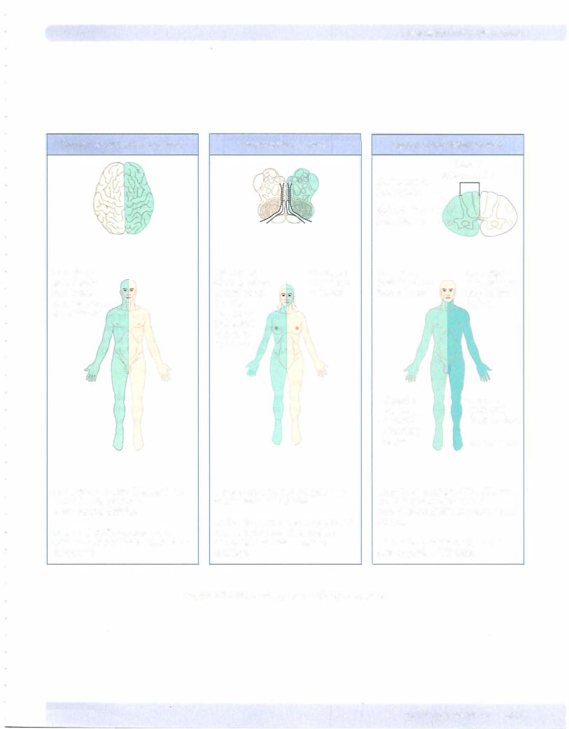
•The brain stem is divided into 3 major subdivisions-the medulla oblongata, pons, and midbrain. The brain stem contains many descending and ascending tracts, the reticular formation, and the sensory and motor cranial nuclei ofCNs Ill-XII. Cranial nerve nuclei forCN Ill and IV are located in the midbrain; cranial nerve nuclei for CN V-Vlll are located in the pons; and cranial nerve nuclei of IX-XII in the medulla. Most ofthe motor nuclei are located medially and the sensory nuclei are located more laterally in the brain stem. Normal functions ofthe cranial nerves and the clinical deficits resulting from lesions ofthe brain stem are listed in Table IV-5-1. Lesions affecting the 3 long tracts to and from the spinal cord will produce contralateral deficits, but lesions ofthe motor or sensory cranial nuclei result in ipsilateral findings.
•The motor nuclei of CNs Ill-VII and IX-XII are lower motor neurons that innervate most ofthe skeletal muscles ofthe head. These lower motor neurons are innervated by upper motor neurons (corticobulbar fibers). The cell bodies of the corticobulbar fibers are found primarily in the motor cortex of the frontal lobe. Corticobulbar innervation of lower motor neurons is primarily bilateral from both the right and left cerebral cortex, except for the innervation ofthe lower facial muscles around the mouth, which are derived only from the contralateral motor cortex. Generally, no cranial deficits will be seen with unilateral corticobulbar lesions, except for drooping ofthe corner of the mouth contralateral to the side of the lesion.
•CN VIII provides sensory pathways for auditory and vestibular systems.
Auditory input depends on the stimulation of hair cells on the organ of Corti due to movement of endolymph within the membranous labyrinth of the inner ear. Axons from the organ of Corti enterthe pons via CN VIII and
synapse in the cochlear nuclei. From the cochlear nuclei, auditory projections bilaterally ascend the brain stem to the superior olivary nuclei, then via the lateral lemniscus to the inferior colliculus, and then to the medial geniculate body of the thalamus. Final auditory projections connect the thalamus with the primary auditory cortex of both temporal lobes. Thus, each auditory cortex receives input from both ears; however, input from the contralateral ear predominates. Lesions of the inner ear or ofthe cochlear nuclei in the
pons will produce total deafness, whereas other lesions central to the cochlear nuclei will primarily affect the ability to localize sound directi(Continued).