Chapter 5 • The Brain Stem
BasilarArtery
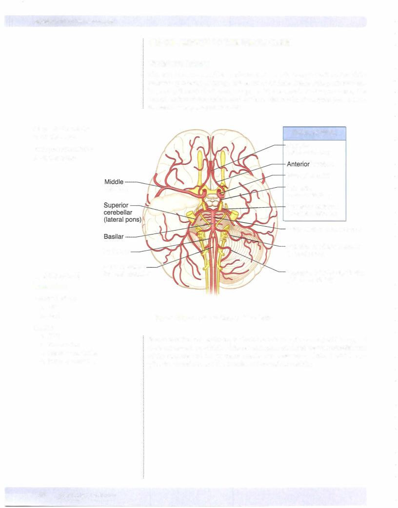
The basilararteryis formed bythejoining ofthe 2 vertebral arteries at the ponto medullary junction. It ascends along the ventral midline ofthe pons and termi nates near the rostral border ofthepons by dividing into the 2 posterior cerebral arteries. Branches include the anterior inferior cerebellar arteries (AICA) and the paramedian arteries.
Branches ofthe basilar artery include: the labyrinthine artery, which follows the course ofthe eighth cranial nerve and supplies the inner ear; the anterior inferior cerebellar artery, which supplies part of the pons and the anterior and inferior regions of the cerebellum; the superior cerebellar artery, which supplies part of the rostral pons and the superior region ofthe cerebellum; and pontine branches, which supply much ofthe pons via paramedian and circumferential vessels.
At the rostral end ofthe midbrain, the basilar artery divides into a pair ofposte rior cerebral arteries. Paramedian and circumferential branches ofthe posterior cerebral arterysupply the midbrain.
BRAIN-STEM LESIONS
There are 2 keys to localizing brain-stem lesions. First, it is uncommon to injure parts ofthe brain stem without involving one or more cranial nerves. The cranial nerve signs will localize the lesion to the midbrain (CN III or IV), upper pons (CN V), lower pons (CN VI, VII, or VIII), or upper medulla (CN IX, X, or XII). Second, ifthe lesion is in the brain stem, the cranial nerve deficits will be seen with a lesion to one or more of the descending or ascending long tracts (cor ticospinal, medial lemniscus, spinothalamic, descending hypothalamic fibers). Lesions in the brain stem to any ofthe long tracts except forthe descending hy pothalamic fibers will result in a contralateral deficit. A unilateral lesion to the descending hypothalamic fibers that results in Horner syndrome is always seen ipsilateral to the side ofthe lesion.
Medial Medullary Syndrome
Medial medullary syndrome is most frequently the result ofocclusion ofthe ver tebral arteryor the anterior spinal artery (Figure IV-5-15). Medial medullary syn dromepresentswith a lesion ofthe hypoglossal nerve as the cranialnerve sign and lesions to both the medial lemniscus and the corticospinal tract. Corticospinal tract lesions produce contralateral spastic hemiparesis ofboth limbs.
Medial lemniscus lesions produce a contralateral deficit of proprioception and touch, pressure, and vibratory sensations in the limbs and body.
Lesions of the hypoglossal nerve in the medulla produce an ipsilateral paralysis ofhalfthe tongue with atrophy. Upon protrusion, the tongue deviatestoward the side ofthe lesion.