Материал: Kaplan USMLE-1 (2013) - Anatomy

Внимание! Если размещение файла нарушает Ваши авторские права, то обязательно сообщите нам

Section IV • Neuroscience

From medialto lateral, the deep cerebellar nuclei in the internal white matter are the fastigial nucleus, interposed nuclei, and dentate nucleus.

Two kinds ofexcitatoryinput enter the cerebellum in the form ofclimbingfibers and mossy fibers. Both types influence the firingofdeep cerebellar nucleibyaxon collaterals.

Climbingfibers originate exclusively from the inferiorolivarycomplexofnuclei on the contralateral side ofthe medulla. Climbing fibers provide a direct power­ ful monosynaptic excitatoryinput to Purkinje cells.

Mossyfibersrepresentthe axonsfromallothersourcesofcerebellarinput. Mossy

fibers provide an indirect, morediffuseexcitatory input to Purkinje cells.

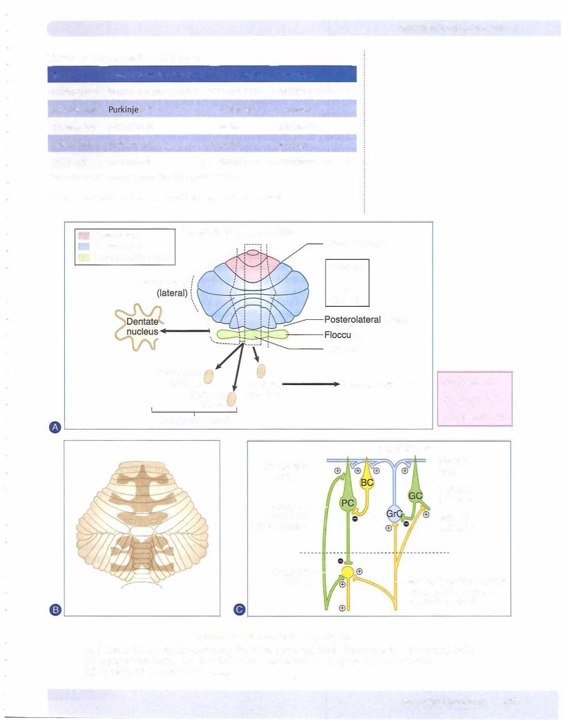
Allmossyfibers exertan excitatory effect on granule cells. Each granule cell sends its axon into the molecular layer, where it gives offcollaterals at a 90-degree angle thatrun parallelto the cortical surface (i.e.,parallelfibers). These granule cell axons stimulate the apical dendrites of the Purkinje cells. Golgi cells receive excitatory input from mossy fibers and from the parallel fibers ofthe granule cells. The Golgi cell in turn inhibits the granule cell, which activatedit in the firstplace.

The basket and stellate cells, which also receive excitatory input from parallel fibers ofgranule cells, inhibit Purkinje cells.

CIRCUITRY

Thebasic cerebellar circuits beginwith Purkinje cells that receiveexcitatoryinput directly from climbing fibers and from parallel fibers ofgranule cells.

Purkinje cell axons project to and inhibit the deep cerebellar nuclei or the ves­ tibular nuclei in an orderlyfashion (Figure IV-6-3).

•Purkinje cells in the flocculonodular lobe project to the lateral vestibular nucleus.

•Purkinje cells in the vermis project to the fastigial nuclei.

•Purkinje cells in the intermediate hemisphere primarily project to the interposed (globose and emboliform) nuclei.

•Purkinje cells in the lateral cerebellar hemisphere project to the dentate nucleus.

Dysfunction

•Hemisphere lesions -7 ipsilateral symptoms: intention tremor, dysmet­ ria, dysdiadochokinesia, scanning dysarthria, nystagmus, hypotonia

•Vermal lesions -7 truncal ataxia

MajorPathway

Purkinje cells -7 deep cerebellar nucleus; dentate nucleus -7 contralateral VL -7 first-degree motor cortex -7 pontine nuclei -7 contralateral cerebellarcortex

424 MEDICAL

Section IV • Neuroscience

Clinical Correlate

Anterior vermis lesions are usually the result of degeneration from alcohol abuse and are present with gait ataxia. Posterior vermis lesions result from medulloblastomas or ependymomas and present with truncal ataxia.

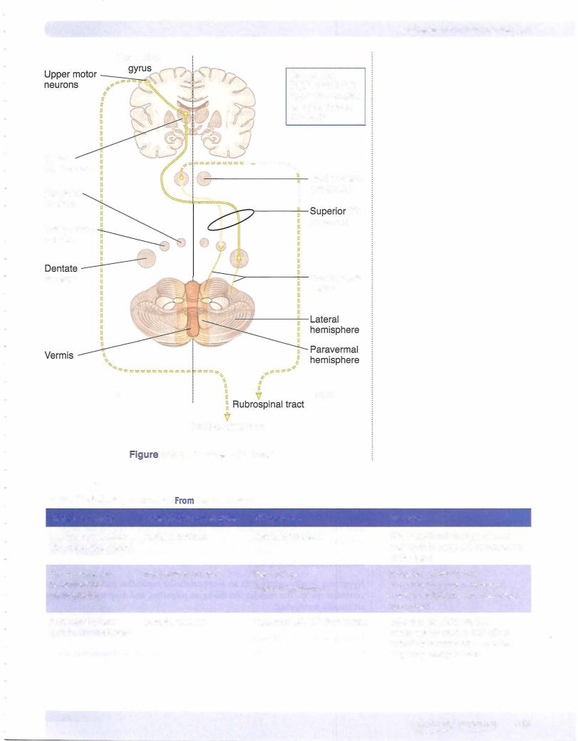
Efferents from the deep cerebellar nuclei leave mainlythrough the SCP and influ­ ence allupper motoneurons. In particular, axons from the dentate and interposed nuclei leave through the SCP, cross the midline, and terminate in the ventrolat­ eral (VL) nucleus ofthe thalamus.

The VL nucleus ofthe thalamus projects to primary motor cortex and influences the firing ofcorticospinal and corticobulbar neurons.

Axons from other deep cerebellar nuclei influence uppermotoneurons in the red nucleus and in the reticular formation and vestibular nuclei.

Cerebellar Lesions

The hallmark of cerebellar dysfunction is a tremor with intended movement without paralysis or paresis. Symptoms associated with cerebellar lesions are ex­ pressed ipsilaterally because the major outflow ofthe cerebellum projects to the contralateral motor cortex, and then the corticospinal fibers cross on their wayto the spinal cord. Thus, unilateral lesions ofthe cerebellum willresult in a patient falling toward the side ofthe lesion.

Lesions that include the hemisphere

Lesions that include the hemisphere produce a number of dysfunctions, mostly involving distal musculature.

An intention tremor is seen whenvoluntarymovements are performed. Forexam­ ple, ifa patient with a cerebellar lesion is asked to pick up a penny, a slight tremor of the fingers is evident and increases as the penny is approached. The tremor is barelynoticeable or is absent at rest.

Dysmetria (past pointing) is the inabilityto stop a movement at the proper place. The patient has difficulty performing the finger-to-nose test.

Dysdiadochokinesia (adiadochokinesia) is the reduced ability to perform alter­ nating movements, such as pronation and supination of the forearm, at a mod­ erately quickpace.

Scanning dysarthria is causedby asynergy ofthe muscles responsible for speech. In scanning dysarthria, patients divide words into syllables, thereby disrupting the melody ofspeech.

Gaze dysfunction occurs when the eyes try to fix on a point: They may pass it or stop too soon and then oscillate a few times before they settle on the target. A nystagmus may be present, particularly with acute cerebellar damage. The nys­ tagmus is often coarse, with the fast component usually directed toward the in­ volved cerebellar hemisphere.

Hypotonia usually occurs with an acute cerebellar insult that includes the deep cerebellar nuclei. The muscles feel flabby on palpation, and deep tendon reflexes are usually diminished.

Lesions to the vermal region

Verma! lesions result in difficultymaintaining posture, gait, or balance (an ataxic gait). Patients with vermal damage maybe differentiated from those with a lesion of the dorsal columns by the Romberg sign. In cerebellar lesions, patients will swayor lose their balancewith their eyes open; in dorsal column lesions, patients sway with their eyes closed.
426 MEDICAL

Источник: https://studfile.net/preview/14638320/