Материал: Kaplan USMLE-1 (2013) - Anatomy
Внимание! Если размещение файла нарушает Ваши авторские права, то обязательно сообщите нам

Section IV • Neuroscience

Open-Angle Glaucoma

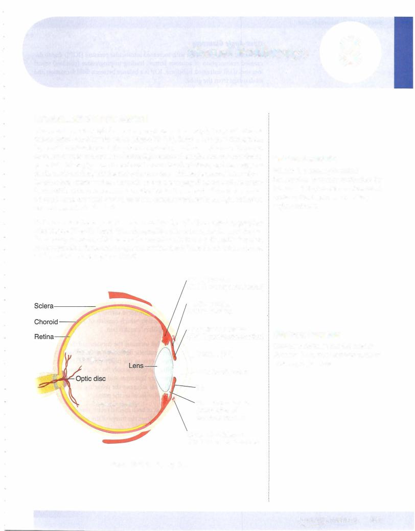
A chronic condition (often with increased intraocular pressure [IOP]) due to de­ creased reabsorption of aqueous humor, leading to progressive (painless) visual loss and, ifleft untreated, blindness. IOP is a balance between fluid formation and its drainage from the globe.

Narrow-Angle Glaucoma

An acute (painful) or chronic (genetic) conditionwith increased IOP due to block­ ade ofthe canal ofSchlemm. Emergencytreatmentprior to surgeryoften involves cholinomimetics, carbonic anhydrase inhibitors, and/or mannitol.

VISUAL REFLEXES

Pupillary Light Reflex

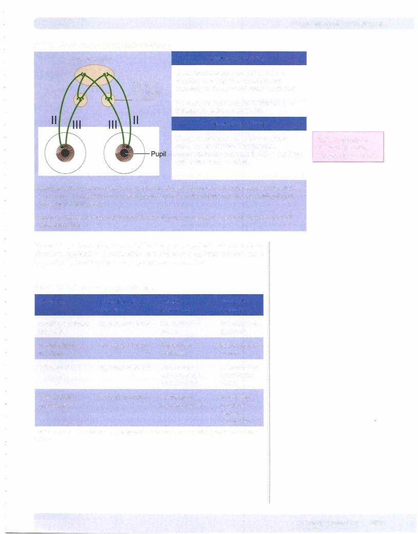
When light is directed into an eye, it stimulates retinal photoreceptors and results in impulses carried in the optic nerve to the pretectal area. Cells in the pretectal area send axons to the Edinger-Westphal nuclei on both sides.

The Edinger-Westphal nucleus is the parasympathetic nucleus ofthe oculomotor nerve and gives rise to preganglionic parasympathetic fibers thatpass in the third cranial nerve to the ciliary ganglion. Because cells in the pretectal area supply both Edinger-Westphal nuclei, shining light into one eye results in constriction of both the ipsilateral pupil (direct light reflex) and contralateral pupil (consensual light reflex).

Accommodation-Convergence Reaction

This reaction occurs when an individual attempts to focus on a nearbyobject after looking at a distant object. The oculomotor nerve carries the efferent fibers from the accommodation-convergence reaction, which consists of3 components: ac­ commodation, convergence, and pupillary constriction.

Accommodation refers to the reflex that increasesthe curvature ofthe lens needed for nearvision. Preganglionic parasympathetic fibers arise in the Edinger-Westphal nucleus and pass via the oculomotor nerve to the ciliary ganglion. Postganglionic parasympathetic fibers from the ciliaryganglionsupplythe ciliarymuscle. Contrac­ tion ofthis muscle relaxes the suspensory ligaments and allows the lens to increase its convexity (become more round). This increases the refractive index ofthe lens, permittingtheimage ofa nearbyobject to focus on the retina.

Convergence results from contraction ofboth medial rectus muscles, which pull the eyes to look toward the nose. This allows the image ofthe near object to focus on the same part ofthe retina in each eye.

Pupillaryconstriction (miosis) results from contraction ofthe constrictor mus­ cle ofthe iris. A smalleraperturegives the optic apparatus agreater depth offield. With Argyll Robertson pupils, both direct and consensual light reflexes are lost, but the accommodation-convergence reaction remains intact. This type ofpupil is often seen in cases ofneurosyphilis; however, it is sometimes seen in patients with multiple sclerosis, pineal tumors, or tabes dorsalis. The lesion site is believed to occur near the pretectal nuclei just rostral to the superior colliculi.

438 MEDICAL
Chapter 8 • Visual Pathways

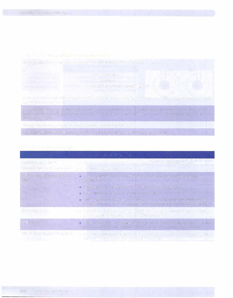
Table IV-8-1. Pupillary Light Reflex Pathway

Pretectal area

Afferent Limb: CN II


Light stimulates ganglion retinal cells


Edinger­

impulses travel up CNll, which projects


bilaterally to the pretectal nuclei (midbrain)


Westphal


nucleus

The pretectal nucleus projects bilaterally


Edinger-Westphal nuclei (CN Ill)


Efferent Limb: CN Ill


Edinger-Westphal nucleus (preganglionic


This is a simplified


parasympathetic) ciliary ganglion

diagram; the ciliary


(postganglionic parasympathetic) pupillary

ganglion is not shown.


sphincter muscle miosis


Because cells in the pretectal area supply the Edinger-Westphal nuclei bilaterally, shining light in one eye constriction in the ipsilateral pupil (direct light reflex) and the contralateral pupil (consensual light reflex).

Because this reflex does not involve the visual cortex, a person who is cortically blind can still have this reflex.

The eye is predominantly innervated by the parasympathetic nervous system. Therefore, application of muscarinic antagonists or ganglionic blockers has a large effect by blocking the parasympathetic nervous system.

Table IV-8-2. Pharmacology ofthe Eye

Structure

Predominant

Receptor

Receptor


Receptor

Stimulation

Blockade

Pupillary sphincter

M3 receptor (PANS)

Contraction

Relaxation

ms. (iris)

a


miosis

mydriasis

Radial dilator

receptor (SANS)

Contraction

Relaxation

ms. (iris)


mydriasis

miosis

Ciliary ms.

M3 receptor (PANS)

Contraction

Relaxation


accommodation

focus for far


for near vision

vision

Ciliary body

receptor (SANS)

Secretion of

Decreased

epithelium


aqueous humor

aqueous


humor


production

Abbreviations: ms., muscle; PANS, parasympathetic nervous system; SANS, sympathetic nervous system

MEDICAL . 439

Section IV • Neuroscience

Table IV-8-3. Accommodation-Convergence Reaction

When an individual focuses on a nearby object after looking at a distant object, 3 events occur:

1.Accommodation

2.Convergence

3.Pupillary constriction (miosis)

In general, stimuli from light visual cortex superior colliculus and pretectal nucleus Edinger-Westphal nucleus (1, 3) and oculomotor nucleus (2).

Accommodation: Parasympathetic fibers contract the ciliary muscle, which relaxes suspensory ligaments, allowing the lens to increase its convexity (become more round). This increases the refractive index ofthe lens, thereby focusing a nearby object on the retina.

Convergence: Both medial rectus muscles contract, adducting both eyes.

Pupillary constriction: Parasympathetic fibers contract the pupillary sphincter muscle miosis.

Table IV-8-4. Clinical Correlates


•

Pupillary Abnormalities

Argyll Robertson pupil

No direct or consensual light reflex; accommodation-convergence intact

(pupillary light-near dissociation)

• Seen in neurosyphilis, diabetes


Relative afferent (Marcus Gunn)


Lesion of afferent limb of pupillary light reflex; diagnosis made with swinging

pupil


flashlight


Shine light in Marcus Gunn pupil pupils do not constrict fully


•

Shine light in normal eye pupils constrict fully


Shine light immediately again in affected eye apparent dilation of both pupils


because stimulus carried through that CN II is weaker; seen in multiple sclerosis

Horner syndrome

Caused by a lesion of the oculosympathetic pathway; syndrome consists of miosis,


ptosis, apparent enophthalmos, and hemianhidrosis

Adie pupil

•

Transtentorial (uncal) herniation


Dilated pupil that reacts sluggishly to light, but better to accommodation; often seen in women and often associated with loss of knee jerks. Ciliary ganglion lesion

Increased intracranial pressure leads to uncal herniation CN Ill compression fixed and dilated pupil, "down-and-out" eye, ptosis

440 MEDICAL

Источник: https://studfile.net/preview/14638320/