Clinical Correlate
Thalamic pain syndrome affects the ventral nuclear group. Patients present with burning, aching pain in contralateral limbs or body. Involvement of the dorsal column
medial lemniscal part ofVPL increases the sensitivity to pain and presents as contralateral loss ofvibratory sense and gait ataxia. Thalamic pain syndrome is resistent to analgesic medications.
Medialnucleargroup (partoflimbicsystem)
Input is from the amygdala, prefrontal cortex, and temporal lobe; output is to the prefrontal cortex and cingulate gyrus. The most important nucleus is the dorso medial nucleus.
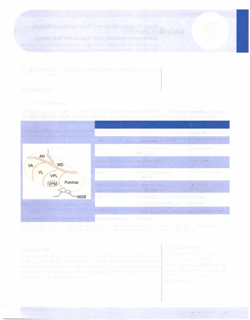
Ventral nucleargroup
MotorVentralNuclanteriori nucleus (VA): Input to VA is from the globus pallidus, substantia nigraVentral. Outputlateralisnucleusto the premotor(VL): and primary motor cortex.
Input to is mainly from the globus pallidus and
VL
the dentate nucleus of the cerebellum. Output is to the primary motor cortex (Brodmann area 4).
SVentralsoryposterolateralNuclei (VPL) nucleus: Input to VPL conveying somatosensory and nociceptive information ascends in the medial lemniscus and spinothalamic
tract. Output is to primary somatosensory cortex (Brodmann areas 3, 1, and 2) ofVentralthe parieposteromedialal lobe. (VPM) nucleus: Input to VPM is from the ascending tri
geminal and taste pathways. Output is to primary somatosensory cortex (Brod
Medialmann argeniculateas 3, 1 , andbody2)(nucleus):ofthe parietal lobe.
Inputis from auditoryinformation that ascends Lateralfrom thegeniculatei ferior colliculusbody (nucleus):. O tput is to primary auditory cortex.
Input is from the optic tract. Output is in the form ofthe geniculocalcarine or visual radiations that project to the primary vi sual (striate) cortex in the occipital lobe.
Midline and lntralaminar Nuclei
Midline and intralarninar nuclei receive input from the brain-stem reticular for mation, and from the spinothalarnic tract. Intralaminar nuclei send pain informa tion to the cingulate gyrus.
These nuclei appear to be important in mediating desynchronization ofthe elec troencephalogram (EEG) duringbehavioral arousal.