Материал: Kaplan USMLE-1 (2013) - Anatomy

Внимание! Если размещение файла нарушает Ваши авторские права, то обязательно сообщите нам

Chapter 8 • Visual Pathways

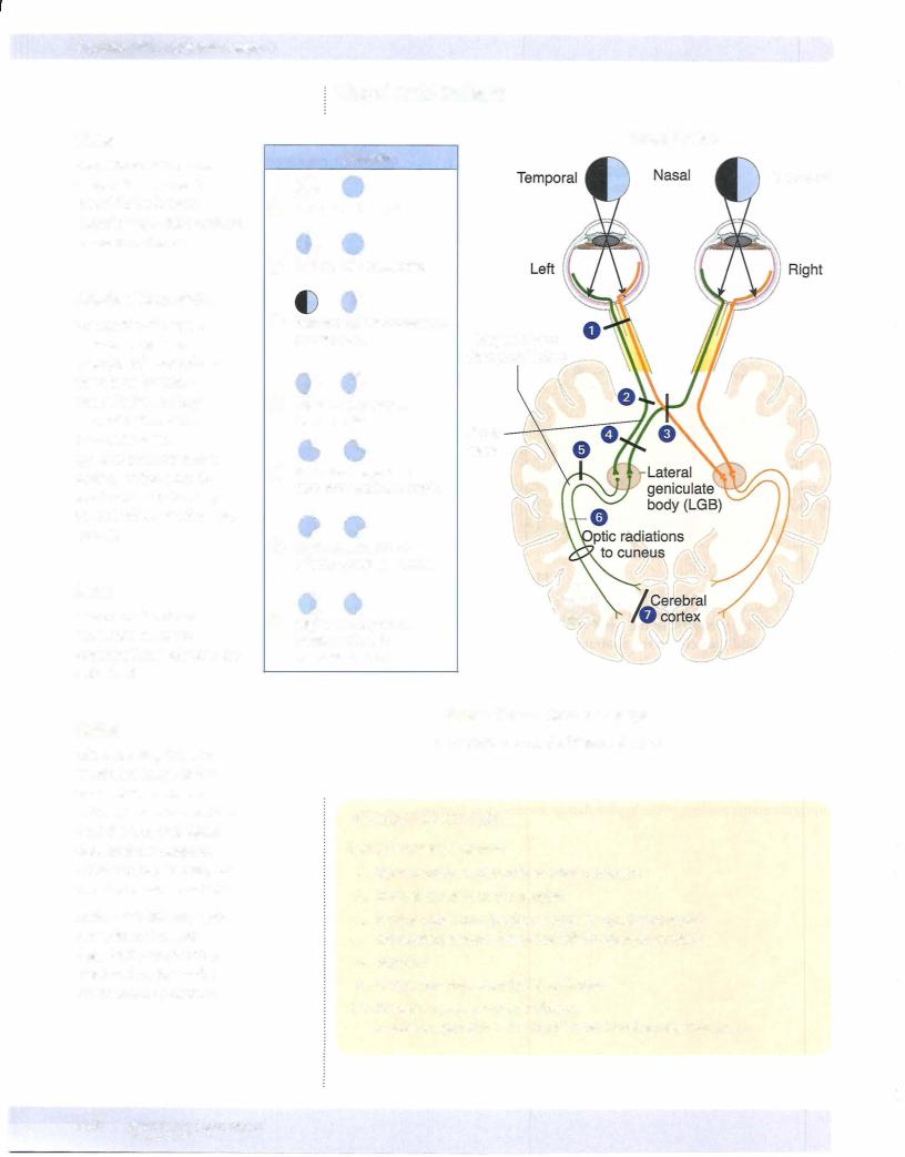
Most fibers from the optic tract project to the lateral geniculate body (LGB); some also project to the pretectal area (light reflex), the superior colliculi (reflex gaze), and the suprachiasmatic nuclei (circadian rhythm). The LGB projects to the primary visual cortex (striate cortex, Brodmann area 1 7) of the occipital lobe via the optic radiations.

•Visual information from the lower retina (upper contralateral visual field) temporal lobe (Meyerloop) lingual gyrus

•Visual information from the upper retina (lower contralateral visual field) parietal lobe cuneus gyrus

The lateral geniculate body (LGB) is a laminated structure thatreceivesinputfrom the optic tract and gives rise to axonsthat terminate on cells in the primaryvisual cortex (striate cortex, Brodmann area 17) ofthe occipital lobe. The LGB laminae maintain a segregation ofinputs from the ipsilateral and contralateral retina.

The axons from the LGB thatproject to the striate cortex are known as optic radia­ tions, visual radiations, or the geniculocalcarine tract. The calcarine sulcus divides the striate cortex (primaryvisual cortex or Brodmann area 17) into the cuneus and the lingual gyri. The cuneus gyrus, which lies on the superior bank ofthe calcarine cortex, receives the medial fibers ofthe visual radiations. The lingual gyrus, which lies on the inferior bank of the calcarine cortex, receives the lateral fibers of the visual radiation. The medial fibers coursing in the visual radiations, which carry input from the upper retina (i.e., the lowercontralateralvisual field), pass from the LGB directlythrough the parietal lobe to reach the cuneus gyrus. Significantly, the lateral fibers coursing in the visual radiations, which carry input from the lower retina (i.e., the upper contralateral visual field), take a circuitous route from the LGB through Meyer loop anteriorly into the temporal lobe. The fibers of Meyer loop then turn posteriorlyand course through the parietal lobe to reach the lingual gyrus in the striate cortex.

-

'

M E D I CAL 443

Section IV • Neuroscience

LESIONS OF THE VISUAL PATHWAYS

Lesions ofthe retina that include destruction of the macula produce a central sco­ toma. The macula is quite sensitive to intense light, trauma, aging, andneurotoxins.

Lesions of an optic nerve produce blindness (anopsia) in that eye and a loss of the sensory limb ofthe light reflex. The pupil ofthe affected eye constricts when light is shined into the opposite eye (consensual light reflex) but not when light is shinedinto the blinded eye (absence ofdirectlight reflex).

Compression ofthe optic chiasm, often the result ofa pituitarytumor ormeningio­ ma, results in a loss ofperipheralvision in bothtemporalfields because the crossing fibers from each nasal retina are damaged. The resultingvisual field defect is called a bitemporal heteronymous hemianopia.

Alllesions past the chiasm produce contralateral defects. Lesions of the optic tract result in a loss of visual input from the contralateral visual field. For ex­ ample, a lesion ofthe right optic tract results in a loss ofinput from the left visual field. This is called a homonymous hemianopia; in this example, a left homony­ mous hemianopia.

Lesions ofthe visual radiations are more commonthan lesions to the optic tract or lateral geniculate body and produce visual field defects (a contralateral homony­ mous hemianopia) similar to those ofthe optic tract ifall fibers are involved.

Lesions restricted to the lateral fibers in Meyer loop, usually in the temporal lobe, result in a loss ofvisual input from the contralateral upper quarter of the visual field. For example, a lesion ofthe temporal fibers in the right visual radiation re­ sults in loss ofvisual input from the upper left quarter ofthe field (a left superior quadrantanopia).

Lesions restricted to the medial fibers in the visual radiation in the parietal lobe result in a loss ofvisual input from the contralateral lower quarter ofthe field (an inferior quadrantanopia).

Lesions inside the primaryvisual cortexare equivalent to those ofthevisualradi­ ations, resulting in a contralateral homonymous hemianopsia, except that macu­ lar (central) vision is spared.

Lesions of the cuneus gyrus are equivalent to lesions restricted to the parietal fibers ofthe visual radiation, with macular sparing.

Lesions ofthe lingula aresimilar to lesions ofthe Meyer's loop fibers exceptfor the presence of macular sparing. The pupillary light reflex is spared in lesions of the radiations or insidevisual cortexbecause fibers ofthepupillarylight reflexleavethe optic tracts to terminate in the pretectal area. The combination ofblindness with intact pupillary reflexes is termed cortical blindness.

444 MEDICAL

Chapter 8 • Visual Pathways

ChapterSummary

•The eyeball is formed by 3 layers: the sclera, choroid, and retina. The shape ofthe lens is modified for near and far vision by the ciliary muscle during the accommodation reflex. The sclera is the external layer and continues anteriorly as the cornea, which is transparent and allows light to enter the eye. The intermediate choroid layer is highly vascularized and pigmented. Anteriorly, the choroid layerforms the ciliary body and iris. The retina contains the photoreceptive layer ofrods (for nightvision and dim light) and cones (for colorvision and high visual acuity).

•The axons ofthe ganglionic cells ofthe retina form the optic nerve at the optic disc.

•The visual pathway is a 3-neuron pathway with the first neuron (bipolar neurons) and the second neuron (ganglionic neurons) located in the retina. The ganglionic axons project from the retina through the optic nerve, optic chiasm, and optic tract to synapse with the third neuron located in the lateral geniculate body ofthe thalamus. These thalamic axons then project via the optic radiations (geniculocalcarine tract) in the parietal lobe to reach the primary visual (striate) cortex at the posterior pole of the occipital lobe. Because the lens inverts images like a camera, each nasal retina receives information from the temporal visual fields, and each temporal retina receives information from the nasal visual field. Atthe optic chiasm, the fibers from the nasal half of each retina decussate while the optic fibers from the temporal half of each retina pass through the chiasm without decussating. Thus, central to the chiasm ipsilateral visual fields project through the contralateral visual pathways. Lesions at the lateral aspect of the optic chiasm produce ipsilateral nasal hemianopsia, whereas midline lesions at the chiasm produce bitemporal heteronymous hemianopsia. Any lesion central to the chiasm results in contralateral homonymous hemianopsia.

•In addition, visual pathways carry optic fibers from the superior and inferior quadrants ofthe visual fields through the retina to the lateral geniculate body. The projections of the superior quadrants to the lower retina reach the lateral geniculate body laterally, synapse, and leave through the lateral

course of Meyer loop in the temporal lobe before rejoining the optic radiation to reach the lower (lingual) gyrus of the striate cortex. Thus, lesions ofthe temporal lobe affecting Meyer loop result in contralateral homonymous superior quadrantanopia. Inferior quadrants ofthe visual fields project to the upper retina and then to the medial aspect ofthe lateral geniculate body. After synapsing in the geniculate body, the axons project completely through the optic radiations to reach the upper (cuneus) gyrus of the striate cortex.

Lesions ofthese more medial fibers produce contralateral homonymous inferior quadrantanopia. Vascular lesions ofthe striate cortex due to occlusion of the posterior cerebral artery result in contralateral homonymous hemianopia with macular sparing.

M EDICAL 445

Источник: https://studfile.net/preview/14638320/