|
DICOM PS3.17 2020a - Explanatory Information |
Page 379 |
Hanging |
Image |
ImageSizeISO Teeth Designation |
Protocol |
Location |
|
(typical) |
Name |
Code |
|
|
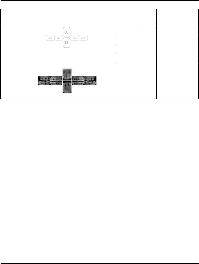
DL-P002E |
01 |
Standard 11, 12, 21, 22 |
|
11 |
|
32, 31, 41, 42 |
|
20 |
Bitewing |
18, 17, 16, 15, 48, 47, 46, |
|
|
|
45 |
|
21 |
|
17, 16, 15, 14 47, 46, 45, |
|
|
|
44 |
Reference: |
23 |
|
27, 26, 25, 24, 37, 36, 35, |
|
|
34 |
|
|
|
|
24 |
|
28, 27, 26, 25, 38, 37, 36, |
|
|
|
35 |
OO.1.2 Ophthalmology
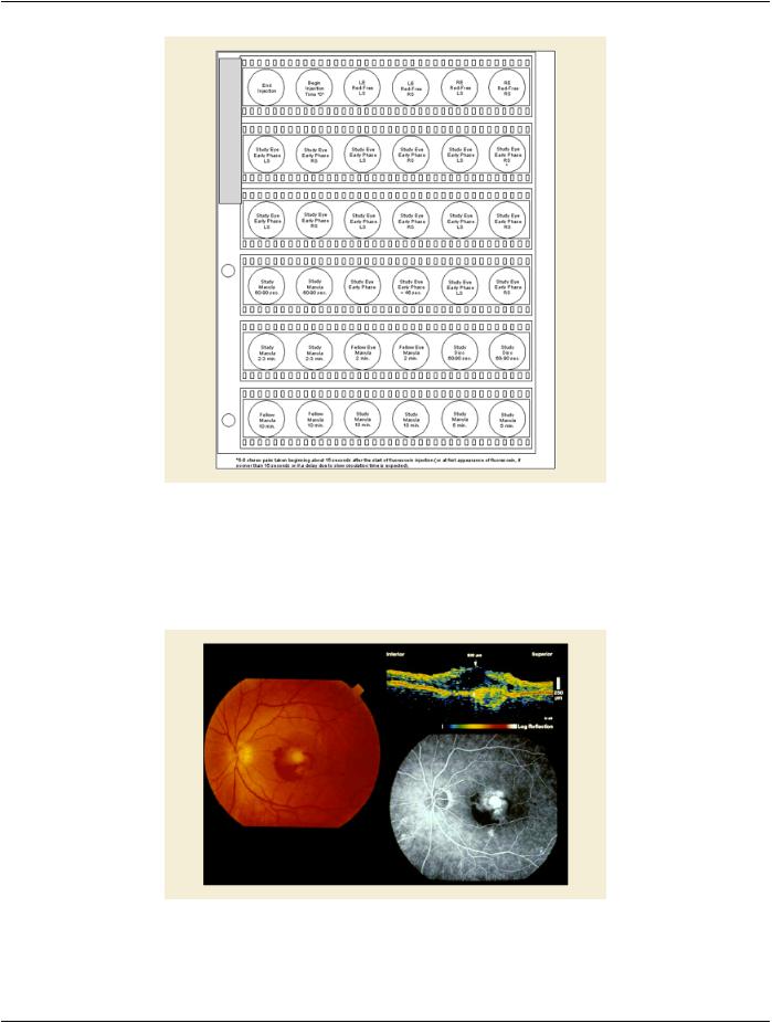
1.A patient in rural Canada visits a general ophthalmologist and is found to have diabetic macular edema. The general ophthalmo- logist would like to discuss the case with a retina specialist before performing laser surgery. A fluorescein angiogram is done with multiple retinal images taken in a timed series after an intravenous injection. The images along with a Structured Display are shared via a Health Information Exchange with a retina specialist in Calgary, who opens them using his Ophthalmology EMR software and consults via phone with the general ophthalmologist. Both physicians view the images in the same layout so the retina specialist can provide accurate guidance for treating the patient.
2.A patient in rural Iowa visits his primary care physician for management of diabetes. Three non-mydriatic (patient's eyes are not dilated) photographs are taken of the back of each eye, and forwarded electronically along with a Structured Display to an oph- thalmologist in Iowa City. The ophthalmologist reads the photos in an agreed upon layout so there is no mistake about what portion of which eye is being viewed. The ophthalmologist is able to tell the primary care physician that his patient does not need to come to Iowa City for face to face ophthalmologic care, but that there is a particular view of the left eye that should be photo- graphed again in 6 months.
 7 Standard + 3 Occlusal Maxilla A Dental Image Layout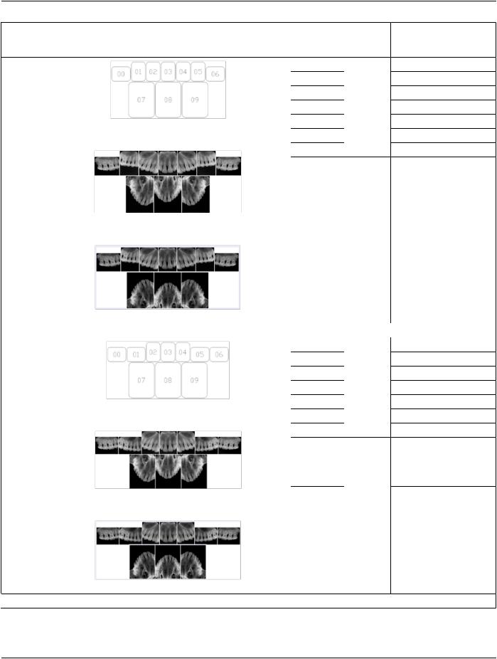
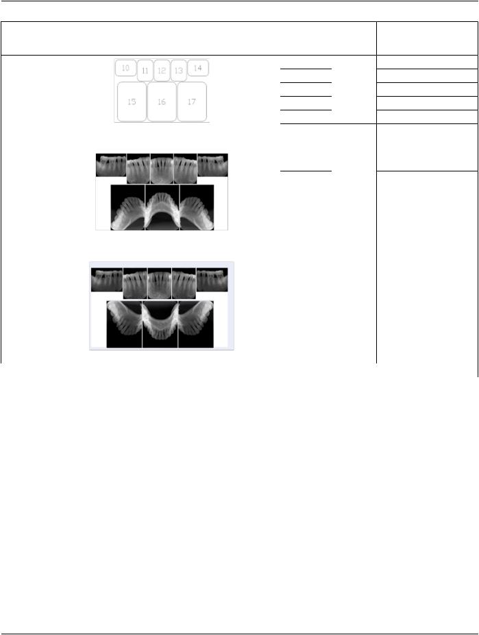
 7 Standard + 3 Occlusal Mandible A Dental Image Layout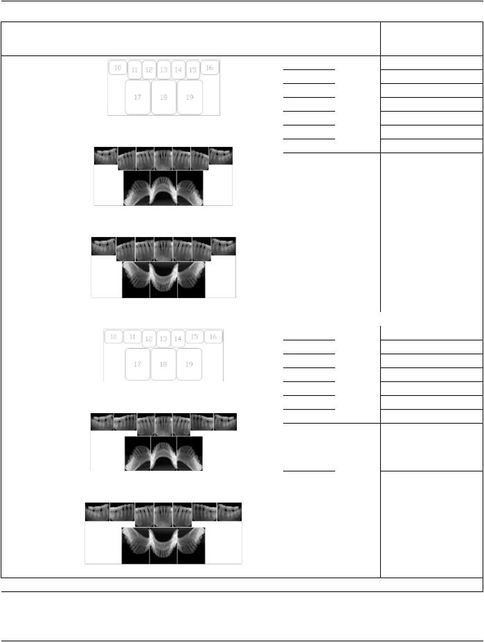
 6 Standard + 4 Bitewing C Dental Image Layout