Page 384 |
DICOM PS3.17 2020a - Explanatory Information |
c.A loop of Rendered Views with a varying observer point
d.A temporal loop of Rendered Views with a varying observer point
Note
Images in this group are not normally measurable because each pixel in the 2D representation may be comprised of data from many pixels in depth along the viewing ray and does not correspond to any particular point in 3D-space.
9.Allow successive display of frames in multi-frame objects in cases 6, 7, and 8.
10.Make size measurements on 2D frames in cases 6, 7, and 8.
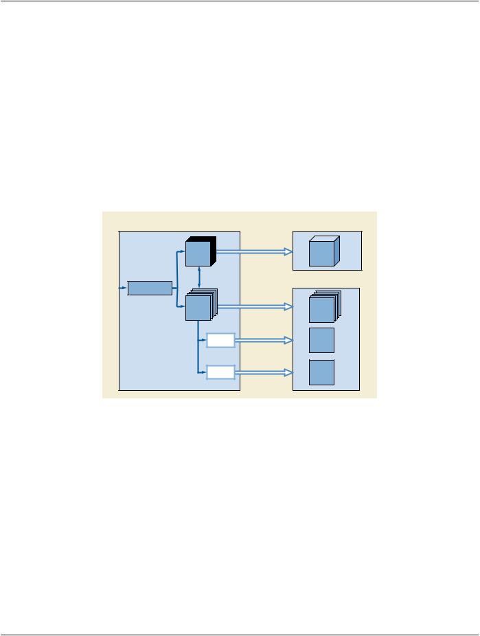
11.Separationofdifferentdatatypesallowsforindependentdisplayand/orprocessingofimagedata(forexample,colorsuppression to expose tissue boundaries, grayscale suppression for vascular flow trees, elastography, etc.)
12.Represent ECG and other physiological waveforms synchronized to acquired images.
13.Two-stage Retrieval: The clinician initially queries for and retrieves all the images in an exam that are directly viewable as sets of frames. Based on the review of these images (potentially on a legacy review application), the clinician may decide to perform advanced analysis of a subset of the exam images. Volume Data sets corresponding to those images are subsequently retrieved and examined.
14.An ultrasound scanner allows user to specify qualitative patient orientation (e.g., Left, Right, Medial, etc.) along with the image data.
15.An ultrasound scanner may maintain a patient-relative frame of reference (obtained such as through a gantry device) along with the image data.
16.Fiducial markers that tag anatomical references in the image data may be specified along with the image data.
17.Key Images of clinical interest are identified and either the entire image, or one or more frames or a volume segmentation within the image must be tagged for later reference.
PP.2.2 Hierarchy of Use Cases
This section organizes the list of use cases into a hierarchy. Section PP.3 maps items in this hierarchy to specific solutions in the DICOM Standard.
1.Data
a.3D Volume Data
i. Static and Dynamic volume data (Cases 1 and 2)
ii. Suitable for applications that create MPR and Render views (Cases 3 and 4)
iii.3D size measurements (Case 5)
b.2D representations of 3D volume data (Cases 6, 7, and 8)
i. Static and Dynamic varieties (Case 9)
ii. 2D size measurements (Case 10)
c.Separation of data types (Case 11)
d.Integrate physiological waveforms with image acquisition (Case 12)
2.Workflow
a.Permit Two-step review (Case 13)
i. Review 2D representations first (potentially on legacy viewer)