Section IV • Neuroscience
COMPONENTS OF THE EAR, AUDITORY, AND VESTIBULAR SYSTEMS
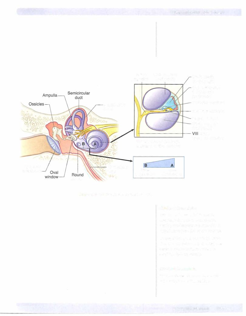
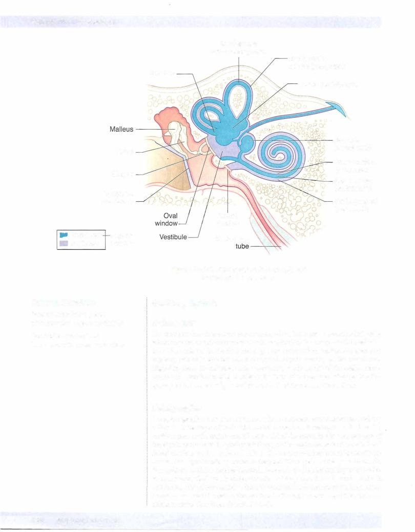
Each ear consists of 3 components: 2 air-filled spaces, the external ear and the middle ear; andthe fluid-filled spaces ofthe inner ear (Figures IV-5-6 and IV-5-7).
Theexternal ear includes thepinna andthe external auditorymeatus, which extends to the tympanic membrane. Soundwavestravelthrough the external auditory canal and cause the tympanic membrane (eardrum) to vibrate. Movement ofthe eardrum causes vibrations ofthe ossicles in the middle ear (i.e., the malleus, incus, and sta pes). Vibrations ofthe ossicles are transferred through the ovalwindow and into the inner ear.
The middle ear lies in the temporal bone, where the chain of 3 ossicles connect the tympanic membrane to the oval window. These auditory ossicles amplify the vibrations received by the tympanic membrane and transmit them to the fluid of the inner ear with minimal energy loss. The malleus is inserted in the tympanic membrane, and the stapes is inserted into the membrane ofthe ovalwindow. Two small skeletal muscles, the tensor tympani and the stapedius, contract to prevent damage to the inner ear when the ear is exposed to loud sounds. The middle-ear cavity communicates with the nasopharynxvia the eustachian tube,which allows air pressure to be equalized on both sides ofthe tympanic membrane.
The inner ear consists ofa labyrinth (osseous and membranous) ofinterconnected sacs (utricle and saccule) and channels (semicircular ducts and the cochlear duct) that contain patches of receptor or hair cells that respond to airborne vibrations or movements ofthe head. Both the cochlear duct and the sacs and channels of the vestibular labyrinth are filled with endolymph, which bathes the hairs ofthe hair cells. Endolymph is unique because it has the inorganic ionic composition ofan intracellular fluid but it lies in an extracellular space. The intracellular ionic composition ofendolymph is important for the function ofhair cells. Perilymph, ionically like a typical extracellular fluid, lies outside the endolymph-filled laby rinth (Figure IV-5-7).