Материал: Kaplan USMLE-1 (2013) - Anatomy

Внимание! Если размещение файла нарушает Ваши авторские права, то обязательно сообщите нам

Section IV • Neuroscience

Auditory Tests

Weber test: place tuning fork on vertex of skull. If unilateral conductive loss vibration is louder in affected ear; if unilateral sensorineural loss vibration is louder in normal ear.

Rinne test: place tuning fork on mastoid process (bone conduction) until vibra­ tion is not heard, then place fork in front of ear (air conduction). If unilateral conductive loss no air conduction after bone conduction is gone; ifunilateral sensorineural loss air conduction present after bone conduction is gone.

VestibularSystem

Sensoryreceptors

The vestibular system contains 2 kinds of sensory receptors, one kind in the utri­ cle and the saccule and the other in the semicircular ducts.

The utricle and the saccule are 2 large sacs, each containing a patch of hair cells in a macula. Each macula responds to linear acceleration and detects positional changes in the head relative to gravity. There are 3 semicircular ducts in the inner ear, each lying in a bony semicircular canal. Each semicircular duct con­ tains an ampullary crest of hair cells that detect changes in angular acceleration resulting from circular movements of the head. The 3 semicircular ducts-an- terior, posterior, and horizontal-are oriented such that they lie in the 3 planes of space. Circular movements of the head in any plane will depolarize hair cells in a semicircular duct in one labyrinth and hyperpolarize hair cells in the cor­ responding duct in the opposite labyrinth.

Vestibularnuclei

There are 4 vestibular nuclei located in the rostral medulla and caudal pons. The vestibular nuclei receive afferents from the vestibular nerve, which innervates re­ ceptors located in the semicircular ducts, utricle, and saccule. Primary vestibular fibers terminate in the vestibular nuclei and the flocculonodular lobe of the cer­ ebellum.

Vestibularfibers

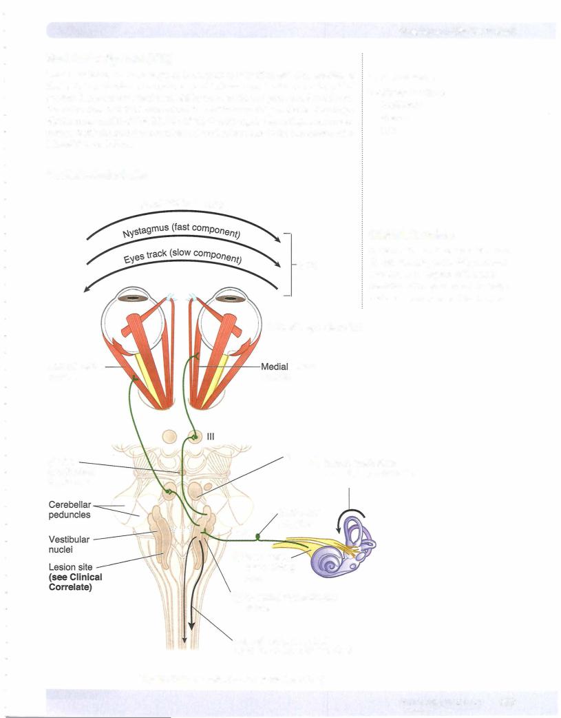
Secondary vestibular fibers, originating in the vestibular nuclei, join the MLF and supply the motor nuclei ofCN III, IV, and VI. These fibers are involved in the pro­ duction ofconjugate eye movements. These compensatory eye movements repre­ sent the efferent limb of the vestibulo-ocular reflex, which enables the eye to re­ main focused on a stationary target during movement of the head or neck. Most of our understanding of the vestibulo-ocular reflex is based on horizontal head turning and a corresponding horizontal movement of the eyes in the direction opposite to that of head turning. For example, when the head turns horizontally to the right, both eyes will move to the left using the following vestibulo-ocular structures. Head turning to the right stimulates hairs cells in the right semicir­ cular ducts. The right eighth nerve increases its firing rate to the right vestibular nuclei. These nuclei then send axons by way of the MLF to the right oculomotor nucleus and to the left abducens nucleus. The right oculomotor nerve to the right medial rectus adducts the right eye, and the left abducens nerve to the left lateral rectus abducts the left eye. The net effect of stimulating these nuclei is that both eyes will look to the left.

402 M EDICAL

Eye MovementControl Systems

For the eyes to move together (conjugate gaze), the oculomotor nuclei and abducens nuclei are interconnected by the medial longitudinal fasciculus (MLF).

Horizontal gaze is controlled by 2 gaze centers: 1 . Frontal eye field (contralateral gaze)

2. PPRF (paramedian pontine reticular formation, ipsilateral gaze)

Nystagmus

Nystagrnus refers to rhythmic oscillations of the eyes slowly to one side followed by a rapid reflex movement in the opposite direction. Nystagmus is defined by the direction of the rapid reflex movement or the fast phase. It is usually horizontal, although rotatory or vertical nystagmus may also occur.

Unilateral vestibular nerve or vestibular nucleus lesions may result in a vestibular nystagmus. In a pathologic vestibular nystagmus, the initial slow phase is the re­ sponse to the pathology, and the fast phase is the correction attempt made bythe cortex in response to the pathology. Consider this example: ifthe left vestibular nerve or nuclei are lesioned, because of the loss of balance between the 2 sides, the right vestibular nuclei are unopposed and act as if they have been stimulated, causing both eyes to look slowly to the left. This is the slow phase of a pathologic vestibular nystagmus. Because the head did not move, the cortex responds by moving both eyes quickly back to the right, the direction of the fast phase of the nystagmus.

Tests for Nystagmus

The integrity of the vestibulo-ocular reflex can be an indicator of brain-stem in­ tegrity in comatose patients. To test this reflex, a vestibular nystagmus is induced byperforming a caloric test in which an examiner introduces warm or cool water into an external auditory meatus. Warm water introduced into the external ear stimulates the horizontal semicircular duct and causes the eyes to move slowly in the opposite direction. Because the head did not turn, the eyes are moved quickly back by the cortex (if intact) toward the same ear where the warm water was introduced, producing a fast phase of nystagmus to the same side. Intro­ duction of cool water into the external ear mimics a lesion; the horizontal duct activity is inhibited on the cool water side, and the opposite vestibular complex moves the eyes slowly toward the cool-water ear. The corrective or fast phase of the nystagrnus moves the eyes quickly away from the ear where the cool water was introduced. A mnemonic which summarizes the direction of the fast phase of vestibular nystagmus in a caloric test toward the warm-water side and away from the cool-water side is COWS; cool, opposite; warm, same.

HORIZONTAL CONJUGATE GAZE

The eyeballs move together in conjugate gaze. The ocular muscles function to move and position both eyes as a unit so that an image falls on a corresponding spot on the retina of each eye. The slightest weakness in the movements of one eye causes diplopia, the presence of a double image, indicating that the image has been shifted to a different position on the retina of the affected side. Although gaze in allplanes is possible, the muscles and cranial nerves involved in horizon­ tal conjugate gaze, or abduction and adduction ofboth eyes together, are the most important eye movements (Figure IV-5- 1 1).
Chapter 5 • The Brain Stem

MEDICAL 405

Section IV • Neuroscience

Abduction ofeach eyeballisperformedlargelybythelateral rectus muscle, which is innervated by the abducens nerve (CN VI). Adduction of the eyeball is per­ formedbythemedialrectusmuscle,whichisinnervatedbythe oculomotornerve (CN III). Therefore, forboth eyes to lookto the right in horizontal gaze, the right abducens nerve and the right lateral rectus muscle must be active to abduct the righteye, and the left oculomotornerve and theleftmedial rectus muscle mustbe active to adduct the left eye. The net effect is that both eyes willlook to the right.

In the brain stem, the abducens nucleus (CN VI) and the oculomotor nucleus (CN III) are situated close to the mid.linejust beneath the fourth ventricle or the cerebral aqueduct, in the pons and rnidbrain. These nuclei are interconnected by thefibers in the MLF. It is the fibers in the MLF thatpermitconjugategaze, either when the target moves or when the head moves, through their interconnections to gaze centers and thevestibularsystem.

Control of Horizontal Gaze

Horizontalgaze is controlledby2 interconnected gaze centers. Onecontrol centeris in the frontal lobe, the frontal eye field (Brodmann area 8). Thisarea acts as a center forcontralateralhorizontal gaze. In the pons is a second gaze center, known as the pontine gaze center or the PPRF, the paramedian pontine reticular formation. This is a center for ipsilateral horizontal gaze. When activatedby neurons in the frontal eye field, the pontine gaze center neurons send axons to synapse with cell bodies in the abducens nucleus, which is actually contained within the pontine gaze center. The pontine gaze center also sends axons that cross immediately and course in the contralateral MLF to reach the contralateral oculomotor nucleus. The net effect of stimulation ofthe left frontal eye field, therefore, is activation ofthe pontine gaze centerontherightanda saccadichorizontal eyemovement ofboth eyes to the right. Horizontal gaze to the right results from activation ofthe right abducens nucleus andthe left oculomotor nucleusbyfibers inthe MLE

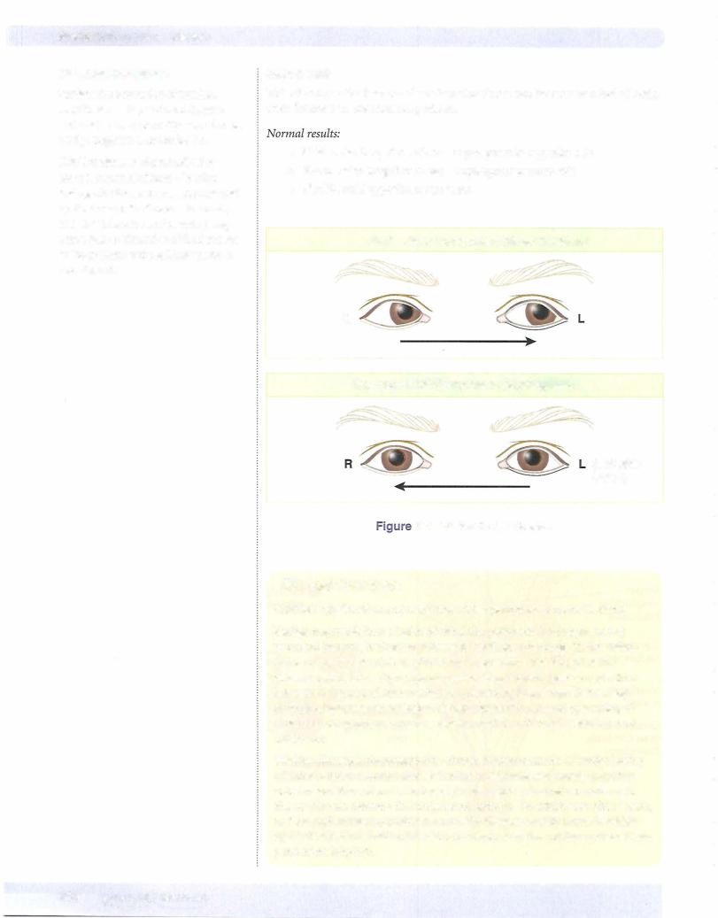
Lesions in the MLF result in an internuclear ophthalmoplegia in which there is an inability to adduct one eye on attempted gaze to the opposite side. For example, a lesion in the right MLF results in an inability to adduct the right eye on an at­ tempted gaze to the left. The left eye abducts normallybut exhibits anystagmus. If the MLF is lesioned bilaterally (as mightbe the case in multiple sclerosis), neither eye adducts on attempted gaze (Figures IV-5-1 1 and IV-5-12), and the abducting eye exhibits a nystagmus.

406 MEDICAL

Источник: https://studfile.net/preview/14638320/