Материал: Oxford American Handbook of Urology ( PDFDrive )

22 CHAPTER 1 Preliminary investigation
Genital symptoms
Scrotal pain
Pathology within the scrotum
•Epididymitis, orchitis, epididymo-orchitis
•Torsion of the testicles
•Torsion of testicular appendages
•Testicular tumor (usually painless)
Referred pain
•Ureteric colic
•Inguinal hernia
•Nerve root irritation or entrapment (ilioinguinal or genitofemoral)
Testicular torsion
Ischemic pain is severe and often accompanied by nausea and vomiting. Torsion presents with sudden onset of pain in the hemiscrotum, sometimes waking the patient from sleep. It may radiate to the groin and/or flank. There is sometimes a history of mild trauma to the testis in the hours before the acute onset of pain.
Similar episodes may have occurred in the past, with spontaneous resolution of the pain (suggesting torsion or spontaneous detorsion). The testis is very tender. It may be high-riding (lying at a higher than normal position in the testis) and may lie horizontally due to twisting of the cord.
Torsion of testicular appendage
Pain is usually not as severe as testicular torsion, and onset can be more gradual, not usually associated with nausea/vomiting.
Epididymitis, orchitis, epididymo-orchitis
These conditions have similar presenting symptoms to those of testicular torsion. Tenderness is most commonly localized to the epididymis. Isolated orchitis is rare today. Untreated epididymitis may involve the testicle secondarily with massive swelling and diffuse tenderness. See p. 33 for advice on attempting to distinguish torsion from epididymo-orchitis.
Testicular tumor
Only 20% of patients present with testicular pain, and most often after minor trauma.
Acute presentations of testicular tumors
•Testicular swelling may occur rapidly (over days or weeks). An associated (secondary) hydrocele is common. A hydrocele, especially in a young person, should always be investigated with an ultrasound to determine whether the underlying testis is normal.
•Rapid onset (days) of testicular swelling can occur. Very rarely, patients present with advanced metastatic disease (high-volume disease in the retroperitoneum, chest, and neck, causing chest, back, or abdominal pain or shortness of breath).

GENITAL SYMPTOMS 23
•Approximately 10–15% of testis tumors present with signs suggesting inflammation (i.e., signs suggesting a diagnosis of epididymo-orchitis—a tender, swollen testis, with redness in the overlying scrotal skin and a fever).
Priapism
Priapism is painful, persistent, prolonged erection of the penis not related to sexual stimulation (see causes in Chapter 12). It can be associated with pharmacological therapies (oral and intracavernosal) for erectile dysfunction. Recurrent or “stuttering” priapism episodes are recurrent but of limited duration. There are two broad categories—low flow (most common) and high flow.
Low-flow (“ischemic”) priapism is due to hematological disease (hemoglobinopathies, sickle cell anemia, thalassemia) or infiltration of the corpora cavernosa with malignant disease, or it is medication related. The corpora cavernosa are very rigid and painful because the corpora are ischemic.
High-flow priapism is due to perineal trauma, which creates an arteriovenous fistula. There is less pain and rigidity.
Diagnosis is usually obvious from the history and examination. Characteristically, the corpora cavernosa are rigid and the glans is flaccid. In low-flow priapism of the erect penis is very tender. Examine the abdomen for evidence of malignant disease and perform a digital rectal examination to examine the prostate. If necessary, corporal blood gas can be used to sort ischemic priapism (pO2 < 30 mmHg, pCO2 > 60 mm Hg, pH < 7.25) from non-ischemic priapism (pO2 > 90 mmHg, pCO2 < 40 mmHg, pH > 7.4).

24 CHAPTER 1 Preliminary investigation
Abdominal examination in urological disease
Because of their retroperitoneal (kidneys, ureters) or pelvic location (bladder and prostate) normal urologic organs are relatively inaccessible to the examining hand when compared with, for example, the spleen or liver. For the same reason, for the kidneys and bladder to be palpable typically implies a fairly advanced disease state in the adult.
It is important that the urologist appreciates the characteristics of other intra-abdominal organs when involved with disease, so that they may be distinguished from urologic organs.
Characteristics and causes of an enlarged kidney
The mass lies in a paracolic gutter, it moves with respiration, is dull to percussion, and can be felt bimanually. It can also be balloted (i.e., bounced, like a ball [balla = “ball” in Italian]) between the hands, one placed on the anterior abdominal wall and one on the posterior abdominal wall.
Common causes of an enlarged kidney
Causes include tumors (renal carcinoma, angiomyolipoma, sarcoma, nephroblastoma), hydronephrosis, pyonephrosis, perinephric abscess, and polycystic disease.
Characteristics and causes of an enlarged liver
The mass descends from underneath the right costal margin; one cannot get above it. It moves with respiration, is dull to percussion, and has a sharp or rounded edge. The surface may be smooth or irregular.
Causes of an enlarged liver
These include infection, congestion (heart failure, hepatic vein obstruc- tion—Budd–Chiari syndrome), cellular infiltration (amyloid), cellular proliferation, space-occupying lesion (polycystic disease, metastatic infiltration, primary hepatic cancer, hydatid cyst, abscess), and cirrhosis.
Characteristics and causes of an enlarged spleen
The mass appears from underneath the costal margin, enlarges toward the right iliac fossa, is firm and smooth, and may have a palpable notch. It is not possible to get above the spleen, it moves with respiration, is dull to percussion, and cannot be felt bimanually.
Causes of an enlarged spleen
These include infections (bacterial, viral infection, protozoal, spirochete); hematological disease; cellular proliferation (leukemias); congestion (portal hypertension due to cirrhosis, portal vein thrombosis, hepatic vein obstruction, congestive heart failure); cellular infiltration; and space-occu- pying lesions (cysts, lymphoma, polycystic disease).

ABDOMINAL EXAMINATION IN UROLOGICAL DISEASE 25
Characteristics of an enlarged bladder
The bladder arises out of the pelvis and is dull to percussion. Pressure of the examining hand may cause a desire to void. In infants and young children it is easier to palpate.
Abdominal distension: causes and characteristics
•Fetus/gravid uterus—very smooth, firm mass, dull to percussion, arising out of the pelvis
•Flatus—hyperresonant (there may be visible peristalsis if the accumulation of flatus is due to bowel obstruction)
•Feces—palpable in the flanks and across the epigastrium, firm and may be identifiable, there may be multiple separate masses in the line of the colon
•Fat
•Fluid (ascites)—fluid thrill, shifting dullness
•Large abdominal masses (massive hepatomegaly or splenomegaly, fibroids, polycystic kidneys, retroperitoneal sarcoma)
The umbilicus and signs and symptoms of associated pathology
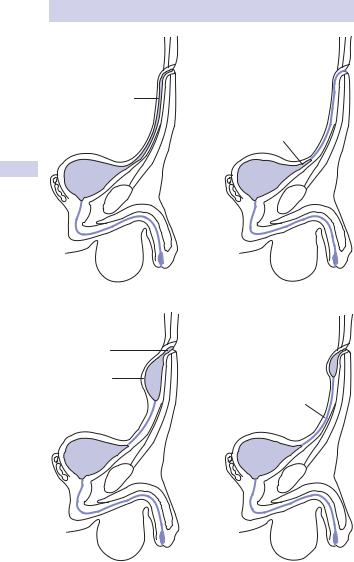
The umbilicus represents the location of four fetal structures—the umbilical vein, two umbilical arteries, and the urachus, which is a tube extending from the superior aspect of the bladder toward the umbilicus. The urachus represents the obliterated vesicourethral canal). The urachus may remain open at various points leading to the following abnormalities (Fig. 1.2):1
•Completely patent urachus communicates with the bladder and leaks urine through the umbilicus. This usually does not present until adulthood because strong contractions of bladder of a child close the mouth of the fistula.
•Vesicourachal diverticulum is a diverticulum in the dome of the bladder. This is usually asymptomatic.
•Umbilical cyst or sinus can become infected, forming an abscess or may chronically discharge infected material from the umbilicus. A cyst can present as an immobile, midline swelling between the
umbilicus and bladder, deep to the rectus sheath. It may have a small communication with the bladder, thus its size can fluctuate as it can become swollen with urine.
Other causes of umbilical masses
These include metastatic deposit (from abdominal cancer, metastatic spread occurring via lymphatics in the edge of the falciform ligament, running alongside the obliterated umbilical vein) and endometriosis (becomes painful and discharges blood at the same time as menstruation).
1 Hinman F Jr (1993). Atlas of Urosurgical Anatomy. Philadelphia: W.B. Saunders.
26 CHAPTER 1 Preliminary investigation
(a) |
(b) |
Patent
urachus
Vesicourachal diverticulum
(c) |
(d) |
Umbilical |
|
sinus |
|
Umbilical |
|
cyst |
Sinus tract |
|
Figure 1.2 Urachal abnormalities. This figure was published in Hinman F Jr, Atlas of Urosurgical Anatomy. Copyright Elsevier 1993.