Материал: Oxford American Handbook of Urology ( PDFDrive )

642 CHAPTER 16 Urological surgery and equipment
Percutaneous nephrolithotomy (PCNL)
Indications
•Stones >3 cm in diameter
•Stones that have failed ESWL and/or an attempt at flexible ureteroscopy and laser treatment
•Staghorn calculi
Preoperative preparation
•CT scan to assist planning the track position and to identify a retrorenal colon1
•Stop aspirin 10 days prior to surgery
•Culture urine (so appropriate antibiotic prophylaxis can be given)
•Cross-match 2 units of blood
•Start IV antibiotics the afternoon before surgery to reduce the chance of septicemia (many stones treated by PCNL are infection stones). If urine is culture negative, use 1.0 g IV ampicillin and IV gentamicin (1.5 mg/kg). Routine antibiotic prophylaxis also reduces the incidence of postoperative UTI.2
Postoperative management
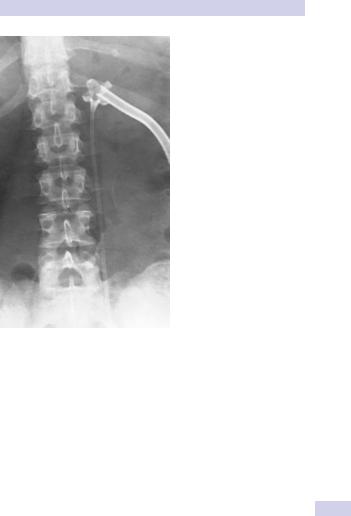
Once the stone has been removed, a nephrostomy tube is left in situ for several days (Fig. 16.15). This drains urine in the postoperative period and tamponades bleeding from the track.
Complications of PCNL and their management
Bleeding
Some bleeding is inevitable, but an amount severe enough to threaten life is uncommon. In most cases it is venous in origin and stops following placement of a nephrostomy tube (which compresses bleeding veins in the track).
If bleeding persists, clamp tube for 10 minutes. If bleeding continues despite this, order urgent angiography, looking for an arteriovenous fistula or pseudoaneurysm, both of which require selective renal artery embolization (required in 1% of PCNLs3) or open exposure of kidney to control bleeding by suture ligation, partial nephrectomy, or nephrectomy.
Septicemia
This occurs in 1–2% of cases. Incidence is reduced by prophylactic antibiotics. Track damage; it is essentially minimal. Cortical loss from track is estimated to be <0.2% of total renal cortex in animal studies.4
1 Hopper KD, Sherman JL, Williams MD, et al. (1987). The variable anteroposterior position of the retroperitoneal colon to the kidneys. Invest Radiol 22:298–302.
2 Inglis JA, Tolly DA (1988). Antibiotic prophylaxis at the time of percutaneous stone surgery. J Endourol 2:59–62.
3 Martin X (2000). Severe bleeding after nephrolithotomy: results of hyperselective embolisation. Eur Urol 37:136–139.

644 CHAPTER 16 Urological surgery and equipment
Damage to the lung and pleura leading to pneumomothorax or pleural effusion can occur with supra-12th rib puncture.
Nephrocutaneous fistula
When the nephrostomy tube is removed from the kidney, a few days after surgery, the 1 cm incision usually closes within 2 days or so.
Occasionally, urine continues to drain percutaneously for a few days and a small stoma bag must be worn. In most of these cases the urine leak will stop spontaneously, but if it fails to do so after a week or so, place a JJ stent to encourage antegrade drainage of urine.
Outcomes
For small stones, the stone-free rate after PCNL is on the order of 90–95%. For staghorn stones, the stone-free rate of PCNL, when combined with postoperative ESWL for residual stone fragments, is on the order of 80–85%.
Procedure-specific consent form—recommended discussion of adverse events
Serious or frequently occurring complications of PCNL
Common
•Temporary insertion of a bladder catheter and ureteric stent or kidney tube needing later removal
•Transient hematuria
•Transient temperature
Occasional
•More than one puncture site may be required
•No guarantee of removal of all stones and need for further operations
•Recurrence of stones
Rare
•Severe kidney bleeding requiring transfusion, embolization, or, at last resort, surgical removal of kidney
•Damage to lung, bowel, spleen, or liver requiring surgical intervention
•Kidney damage or infection needing further treatment
•Overabsorption of irrigating fluids into the blood system, causing strain on heart function
Alternative treatment includes external shock wave treatments, open surgical removal of stones, observation.
This page intentionally left blank

646 CHAPTER 16 Urological surgery and equipment
Ureteroscopes and ureteroscopy
Instruments
Two types of ureteroscope are in common use—the semi-rigid ureteroscope and the flexible ureteroscope.
Semi-rigid ureteroscopes
These instruments have high-density fiber-optic bundles for light (noncoherently arranged) and image transmission (coherently arranged to maintain image quality). For equivalent light and image transmission using glass rod lenses, thicker lenses are required than with fiber-optic bundles.
Consequently, semi-rigid ureteroscopes can be made smaller, while maintaining the size of the instrument channel. In addition, the instrument can be bent by several degrees without the image being distorted.
The working tip of most current models is on the order of 7 to 8 Fr., with the proximal end of the scope being on the order of 11 to 12 Fr. There is usually at least one working channel of at least 3.4 Fr.
Flexible ureteroscopes
The fiber-optic bundles in flexible ureteroscopes are the same as those in semi-rigid scopes, only of smaller diameter. Thus, image quality and light transmission are not as good as with semi-rigid scopes, but are usually adequate.
The working tip of most current models is on the order of 7 to 8 Fr., with the proximal end of the scope being on the order of 9 to 10 Fr. There is usually at least one working channel of at least 3.6 Fr.
The great advantage of the flexible ureteroscope over the semi-rigid variety is the ability to perform controlled deflection of the end of the scope (active deflection). Behind the actively deflecting tip of the scope is a segment of the scope that is more flexible than the rest of the shaft. This section is able to undergo passive deflection—when the tip is fully actively deflected, by advancing the scope further, this flexible segment allows even more deflection.
Flexible ureteroscopes have recently been developed that have two actively deflecting segments.
Flexible ureteroscopes are intrinsically more intricate and are therefore less durable than semi-rigid scopes.
Ureteroscopic irrigation systems
Normal saline is used (high-pressure irrigation with glycine or water would lead to fluid absorption from pyelolymphatic or venous backflow). Irrigation by gravity pressurization alone (the fluid bag suspended above the patient without any applied pressure) will produce flow that is inadequate for visualization because the long, fine-bore irrigation channels of modern ureteroscopes are inherently high resistance.
Several methods are available—hand-inflated pressure bags, foot pumps, and hand-operated syringe pumps. Whatever system is chosen, use the minimal flow required to allow a safe view so as to avoid flushing the stone out of the ureter and into the kidney; you may not be able to retrieve it from there.