Материал: Oxford American Handbook of Urology ( PDFDrive )
BLADDER CANCER: URINARY DIVERSION AFTER CYSTECTOMY 267
This orthotopic neobladder should require no catheter, unless the pouch is too large and fails to empty adequately. In this case, the patient must be prepared to perform intermittent self-catheterization. Initially, bladder irrigations once or twice a day are necessary because of mucous production that gradually diminishes with time.
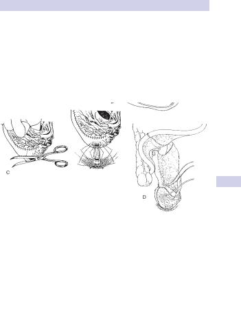
Popular ileal pouches include those of Studer (Fig. 6.7), Camey II, and Kock; ileocecal pouches include the Indiana and Mainz I. The choice of pouch often comes down to the surgeon’s preference; they all carry similar complication risks.
a
b
c
d
Figure 6.7 (a) The distal 40–44 cm of resected ileum opened along the antimesenteric border with scissors. Spatulated ureters are anastomosed end to side with 4–0 running suture on either side of proximal end of afferent tubular ileal limb. Ureters are stented. (b) The two medial borders of the U-shaped, opened, distal ileal segment are oversewn with a single-layer seromuscular continuous suture. The bottom of the U is folded between the two ends of the U. (c) Before complete closure of the reservoir, an 8–10 mm hole is cut into the most caudal part of the reservoir (left). Six sutures are placed between the seromuscular layer of the anastomotic area of the reservoir and the membranous urethra (right). An 18 Fr urethral catheter is inserted. (d) Before complete closure of the pouch, a cystostomy tube is inserted and brought out suprapubically adjacent to the wound. Reprinted with permission from Studer UE, Danuser H, Hochreiter W, et al. (1996). World J Urol 14(1):29–39.

268 CHAPTER 6 Urological neoplasia
Previously irradiated bowel can be cautiously used to form pouches, though complications are more likely. In orthotopic neobladders, incontinence is more common in men than in women, and women have a higher likelihood of being in retention and requiring long-term intermittent catheterization.
Complications relating to continent diversions and neobladders
•Urinary leakage and peritonitis
•Pelvic abscess
•Stone formation
•Catheterizing difficulties and stomal stenosis
•Urinary incontinence and nocturnal enuresis
•Pouch-ureteral reflux and UTI
•Uretero-pouch anastomotic stricture
•Late neobladder rupture
Metabolic abnormalities
These include early fluid and electrolyte imbalances. Later, urinary electrolyte absorption may cause hyperchloremic acidosis, and loss of the distal small bowel (distal 15 cm of ileum) may result in vitamin B12 deficiency. Metabolic acidosis is less likely in patients with normal renal function; treatment is with sodium bicarbonate and potassium citrate.
Annual B12 and CBC monitoring should be undertaken, with supplementation if necessary.
Adenocarcinoma
Adenocarcinoma may develop (5%) in diversions that involve a segment of colon. While more likely with ureterosigmoidostomy with constant contact of feces with the urinary stream, the cause is the carcinogenic bacterial metabolism of urinary nitrosamines.
This tends to occur near the inflow of urine. It is therefore advisable to perform annual visual surveillance of colon urinary diversions after 10 years. If the urethra is in situ, as with orthotopic neobladder, annual urethroscopy and cytology are suggested.
This page intentionally left blank

270 CHAPTER 6 Urological neoplasia
Transitional cell carcinoma (UC) of the renal pelvis and ureter
UC accounts for 90% of upper urinary tract tumors, the remainder being benign inverted papilloma, fibroepithelial polyp, squamous cell carcinoma (associated with longstanding calculus disease), adenocarcinoma (rare), and various rare nonurothelial tumors, including sarcoma.
•Renal pelvic UC is uncommon, accounting for 10% of renal tumors and 4% of all UC.
•Ureteral UC is rare, accounting for only 1% of all newly presenting UC. Half are multifocal; 75% are located distally; while only 3% are located in the proximal ureter.
Risk factors are similar to those for UC in the bladder (see p. 246).
•Males are affected three times more than females.
•Incidence increases with age.
•Smoking confers a two-fold risk, and there are various occupational causes.
UC does not have a genetic hereditary form, although there is a high incidence of upper tract UC in families from some villages in Balkan countries (Balkan nephropathy) that remains unexplained.
Pathology and staging
The tumor usually has a papillary structure, but occasionally it is solid. It is bilateral in 2–4%. It arises within the renal pelvis, less frequently in one of the calyces or ureter. Histologically, features of UC are present, described below. Staging is by the TNM classification. Spread is by
•Direct extension, including into the renal vein and vena cava
•Lymphatic spread to para-aortic, paracaval, and pelvic nodes
•Blood-borne spread, most commonly to liver, lung, and bone
Presentation
•Painless hematuria (80%)
•Flank pain (30%), often caused by clots passing down the ureter (“clot colic”)
•Associated with synchronous bladder UC (4%)
•At follow-up, ~50% of patients will develop a metachronous bladder UC and 2% will develop contralateral upper tract UC
Investigations
Diagnosis is usually made on urine cytology and IVP or CTU, respectively, revealing malignant cells and a filling defect in the renal pelvis or ureter. If doubt exists, selective ureteral urine cytology, retrograde pyeloureterography, or flexible ureteroscopy are indicated.
If ultrasound and cystoscopy are normal during the investigation of hematuria, an IVP or CT is recommended. Staging imaging is obtained by contrast-enhanced abdominal CT, chest X-ray, and, occasionally, isotope bone scan.

TRANSITIONAL CELL CARCINOMA OF RENAL PELVIS AND URETER 271
Staging is by the TNM (2002) classification (see Table 6.10) following histological confirmation of the diagnosis. All cases rely on physical examination and imaging, the pathological classification corresponding to the TNM categories See Box 6.4 for corresponding 5-year survival rates.
Treatment and prognosis
Nephroureterectomy
If staging indicates nonmetastatic disease in the presence of a normal contralateral kidney, the gold-standard treatment with curative intent is nephroureterectomy, open or laparoscopic.
The open approach uses either a long transperitoneal midline incision or separate flank and iliac fossa incisions. The entire ureter is taken with a cuff of bladder, because of the 50% incidence of subsequent ureteral stump recurrence. Follow-up should include annual cystoscopy and IVP or CTU to detect metachronous UC development.
Table 6.10 TNM staging of carcinomas of the renal pelvis and ureter
Tx |
Primary tumor cannot be assessed |
|
|
T0 |
No evidence of primary tumor |
|
|
Ta |
Noninvasive papillary carcinoma |
|
|
Tis |
Carcinoma in situ |
|
|
T1 |
Tumor invades subepithelial connective tissue |
|
|
T2 |
Tumor invades muscularis propria |
|
|
T3 |
Tumor invades beyond muscularis propria into perinephric or |
|
|
|
periureteral fat or renal parenchyma |
|
|
T4 |
Tumor invades adjacent organs or through kidney into perinephric fat |
|
|
Nx |
Regional (para-aortic) lymph nodes cannot be assessed |
|
|
N0 |
No regional lymph node metastasis |
|
|
N1 |
Metastasis in a single lymph node <2 cm |
|
|
N2 |
Metastasis in a single lymph node 2–5 cm or multiple nodes <5 cm |
|
|
N3 |
Metastasis in a single lymph or multiple nodes >5 cm |
|
|
Mx |
Distant metastasis cannot be assessed |
|
|
M0 |
No distant metastasis |
|
|
M1 |
Distant metastasis present |
|
|
|
|
|
|
Box 6.4 5-year survival
• |
Organ-confined disease |
T1,2 |
60–100% |
• |
Locally advanced |
T3,4 |
20–50% |
• |
Node-positive disease |
N+ |
15% |
• |
Pulmonary, bone metastases |
M+ |
10% |