Материал: part17

Внимание! Если размещение файла нарушает Ваши авторские права, то обязательно сообщите нам

DICOM PS3.17 2020a - Explanatory Information​

Page 501​

0.0033s

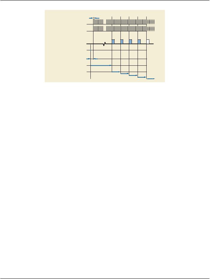
ECG Lead 1 sampling at 300Hz

ECG Lead 2 sampling at 300Hz

time

Nominal start time of

ECG Acquisition Time

0.01s

Start ECG sampling

1s

Start time of Image Acquisition

0.033s

Multi-frame Image Acquisition, at 30fps

Figure FFF.2.1-2. Example of ECG Recording at Acquisition Modality​

FFF.2.1.1.4.1 Enhanced XA Image Without Cardiac Synchronization​

In this example, the Enhanced XA image does not contain information of the cardiac cycle phases.​

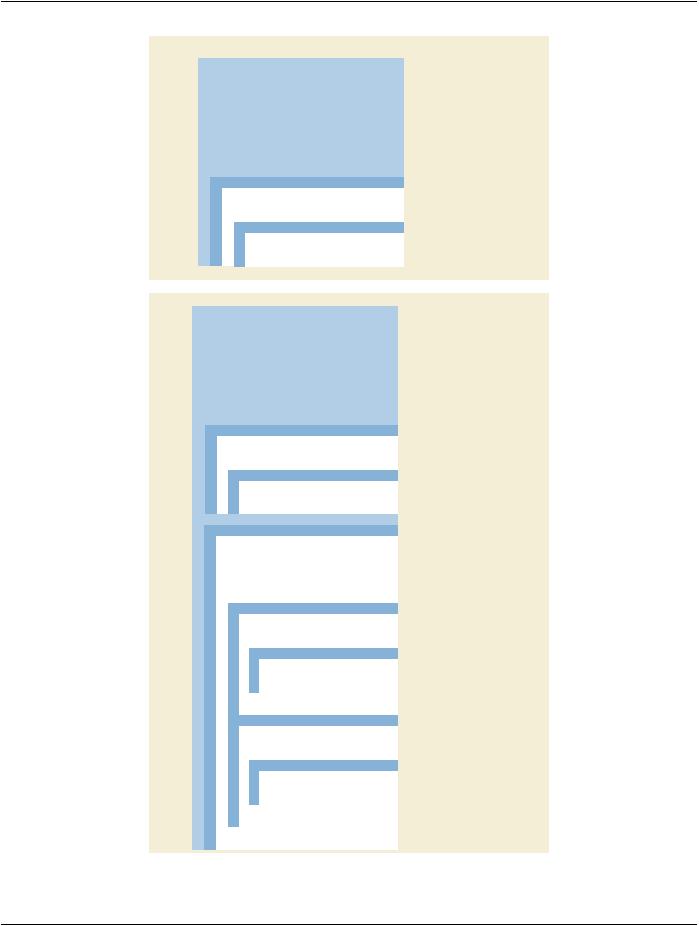
The Attributes that define the two different SOP Instances (Enhanced XA and General ECG) of this example are described in Fig-​ ure FFF.2.1-3.​

Enhanced XA SOP Instance​

- Standard -​

Page 502​

DICOM PS3.17 2020a - Explanatory Information​

Enhanced XA SOP Instance


= UID “A”

Study Instance UID

(0020,000D)


...


Series Instance UID

(0020,000E)

= UID “B”


Modality

(0008,0060)

= XA


...


Synchronization Frame of Reference UID

(0020,0200)

= UID “C”

Synchronization Trigger

(0018,106A)

= NO TRIGGER

Acquisition Time Synchronized

(0008,1800)

= Y

...


SOP Instance UID

(0008,0018)

= UID “D”

Referenced Instance Sequence

(0008,114A)


Item 1


>Referenced SOP Class UID

(0008,1150)

= 1.2.840.10008.5.1.4.1.1.9.1.2


>Referenced SOP Instance UID

(0008,1155)

= UID “E”


>Purpose of Reference Code Sequence

(0040,A170)


Item 1


= 121303


>>Code Value

(0008,0100)


= DCM


>>Coding Scheme Designator

(0028,9503)


= Simultaneous ECG


>>Code Meaning

(0008,0104)

General ECG SOP Instance


= UID “A”

Study Instance UID

(0020,000D)


...


Series Instance UID

(0020,000E)


= UID “F”


Modality

(0008,0060)


= ECG


...


Synchronization Frame of Reference UID

(0020,0200)


= UID “C”

Synchronization Trigger

(0018,106A)


= NO TRIGGER

Acquisition Time Synchronized

(0008,1800)


= Y

...


SOP Instance UID

(0008,0018)


= UID “E”

Referenced Instance Sequence

(0008,114A)


Item 1


>Referenced SOP Class UID

(0008,1150)

= 1.2.840.10008.5.1.4.1.1.12.1.1


>Referenced SOP Instance UID

(0008,1155)


= UID “D”


>Purpose of Reference Code Sequence

(0040,A170)


Item 1


= 121316


>>Code Value

(0008,0100)


>>Coding Scheme Designator

(0028,9503)


= DCM


= Image related to standalone object


>>Code Meaning

(0008,0104)


Waveform Sequence

(5400,0100)


Only one ECG Multiplex Group


Item 1


>Multiplex Group Time Offset

(0018,1068)

= 10


= ORIGINAL


>Waveform Originality

(003A,0004)


>Number of Waveform Channels

(003A,0005)

= 2


>Number of Waveform Samples

(003A,0010)

= 1500


= 300


>Sampling Frequency

(003A,001A)


>Channel Definition Sequence

(003A,0200)


First ECG Channel


Item 1


= 1


>>Waveform Channel Number

(003A,0202)


= ECG Recording 1


>>Channel Label

(003A,0203)


>>Channel Source Sequence

(003A,0208)


Item 1


= 2:1


>>Code Value

(0008,0100)


= MDC


>>Coding Scheme Description

(0008,0102)


= Lead I


>>Code Meaning

(0008,0104)


= 0.0


>>Channel Time Skew

(003A,0214)


= 16


>>Waveform Bits Stored

(003A,021A)


Item 2


= 2


>>Waveform Channel Number

(003A,0202)


= ECG Recording 2


>>Channel Label

(003A,0203)


>>Channel Source Sequence

(003A,0208)


Item 1


= 2:2


>>Code Value

(0008,0100)


= MDC


>>Coding Scheme Description

(0008,0102)


= Lead II


>>Code Meaning

(0008,0104)


>>Channel Time Skew

(003A,0214)

= 0.0


>>Waveform Bits Stored

(003A,021A)

= 16


>Waveform Bits Allocated

(5400,1004)

= 16


>Waveform Sample Interpretation

(5400,1006)


= SS


Figure FFF.2.1-3. Attributes of ECG Recording at Acquisition Modality​

- Standard -​

DICOM PS3.17 2020a - Explanatory Information​

Page 503​

FFF.2.1.1.4.2 Enhanced XA Image With Cardiac Synchronization​

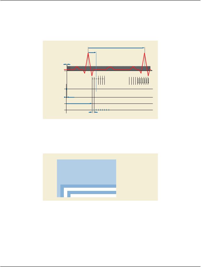
In this example, the heart rate is 75 beats per minute. As the image is acquired during a period of four seconds, it contains five​ heartbeats.​

The ECG signal is analyzed to determine the R-peaks and to relate them to the angiographic frames. Thus the Enhanced XA image​ contains information of this relationship between the ECG signal and the frames.​

ECG Lead 1 sampling at 300Hz

R

800ms

R

90ms


3.3ms


time

Image time

Nominal start time of

ECG Acquisition Time

Frame “i” Reference Time

10ms

Start ECG sampling

1s

Start time of Image Acquisition

Multi-frame Image Acquisition, at 30fps

33ms

Figure FFF.2.1-4. Example of ECG information in the Enhanced XA image​

The Attributes that define the two different SOP Instances (Enhanced XA and General ECG) of this example are described in the​ figures of the previous example, in addition to the Attributes described in Figure FFF.2.1-5.​

Enhanced XA SOP Instance​

Enhanced XA SOP Instance


= REALTIME

Cardiac Synchronization Technique

(0018,9037)


= ECG

Cardiac Signal Source

(0018,9085)


Cardiac RR Interval Specified

(0018,9070)

= 800


Intervals Acquired

(0018,1083)

= 5


Intervals Rejects

(0018,1084)

= 0


...


Per-Frame Functional Groups Sequence

(5200,9230)


...


(0008,114A)


Item 1


Frame “i”


>Cardiac Synchronization Sequence

(0018,9118)


Item 1


>>Nominal Cardiac Trigger Delay Time

(0020,9153)

= 90


Figure FFF.2.1-5. Attributes of Cardiac Synchronization in ECG Recording at Acquisition Modality​

FFF.2.1.2 Multi-modality Waveform Synchronization​

TheseapplicationcasesarerelatedtotheresultsofanX-RayacquisitionandsimultaneousECGdatarecordingondifferentequipment.​ The concepts of synchronized time and triggers are involved.​

The two modalities may share references on the various entity levels below the Study, i.e., Series and Image UID references using​ non-standard mechanisms. Nothing in the workflow requires such references. For more details about UID referencing, refer to the​ previous application case "ECG Recording at Acquisition Modality" (see Section FFF.2.1.1).​

- Standard -​

Page 504​

DICOM PS3.17 2020a - Explanatory Information​

If both modalities share a common data store, a dedicated post-processing station can be used for combined display of waveform​ and image information, and/or combined functional analysis of signals and pixel data to time relate the cardiac cycle phases to the​ angiographic frames. The storage of the waveform data and images to PACS or media will preserve the combined functional capab-​ ilities.​

In these application cases, this post-processing activity is outside the scope of the acquisition modalities. For more details about the​ relationship between cardiac cycle and angiographic frames, refer to the previous application case "ECG Recording at Acquisition​ Modality" (see Section FFF.2.1.1).​

FFF.2.1.2.1 Both Modalities Synchronized Via NTP​

FFF.2.1.2.1.1 User Scenario​

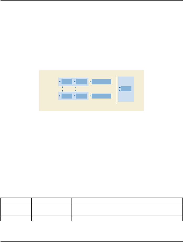
Image runs are taken by the image acquisition modality. Waveforms are recorded by the waveform acquisition modality. Both modal-​ ities are time synchronized via NTP. The time server may be one of the modalities or an external server. The resulting objects will​ include the time synchronization concept.​


Imaging Modality


Workstation


X-Ray


DICOM


DICOM Enhanced XA


Acquisition


Encoding


SOP Instance


Patient


References

Time


Application


UID

Synchronized


(Viewer…)


ECG


DICOM


DICOM General ECG


Acquisition


Encoding


SOP Instance


Waveform Modality


Network,


Offline Media

Figure FFF.2.1-6. Scenario of Multi-modality Waveform Synchronization​

FFF.2.1.2.1.2 Encoding Outline​

Dedicated Waveform IODs exist to store captured waveforms. In this case, General ECG IOD is used to store the waveform data.​

Depending on the degree of coupling of the modalities involved, the usage of references on the various entity levels can vary. While​ there is a standard DICOM service to share Study Instance UID between modalities (i.e., Worklist), there are no standard DICOM​ services for sharing references below the Study level, so any UID reference to the Series and Image levels is shared in a proprietary​ manner.​

With the Synchronization Module information, the method to implement the common time-base can be documented.​

The Enhanced XA IOD provides a detailed "per frame" timing to encode timing information related to each frame.​

FFF.2.1.2.1.3 Encoding Details​

This section provides detailed recommendations of the key Attributes to address this particular scenario.​

FFF.2.1.2.1.3.1 Enhanced XA Image​

Table FFF.2.1-12. Enhanced X-Ray Angiographic Image IOD Modules​

IE​

Module​

PS3.3 Reference​

Usage​

Frame of Reference​Synchronization​

C.7.4.2​

Specifiesthattheimageacquisitionistimesynchronized​


with the ECG acquisition. Will have the same content as​


the General ECG SOP Instance.​

Image​

Enhanced XA/XRF Image​ C.8.19.2​

Specifies the date and time of the image acquisition.​

- Standard -​


DICOM PS3.17 2020a - Explanatory Information​

Page 505​

Table FFF.2.1-13. Enhanced XA Image Functional Group Macros​


Functional Group Macro​


PS3.3 Reference​

Usage​


Frame Content​

C.7.6.16.2.2​

Provides timing information to correlate each frame to any​


externally recorded waveform.​


FFF.2.1.2.1.3.1.1 Synchronization Module Recommendations​


This Module is used to document the synchronization of the two modalities.​


Table FFF.2.1-14. Synchronization Module Recommendations​


Attribute Name​


Tag​

Comment​


SynchronizationFrameofReferenceUID​

(0020,0200)​

The UTC Synchronization UID "1.2.840.10008.15.1.1" is used​


in this case.​


Synchronization Trigger​


(0018,106A)​

The value "NO TRIGGER" is used for the case of time​


synchronization via NTP.​


Acquisition Time Synchronized​


(0018,1800)​

The value "Y" is used in this scenario.​


Time Source​


(0018,1801)​

The same value as in the related General ECG SOP Instance​


is used in this scenario.​


Time Distribution Protocol​


(0018,1802)​

The value "NTP" is used in this scenario.​


NTP Source Address​


(0018,1803)​

The same value as in the related General ECG SOP Instance​


is used in this scenario.​


FFF.2.1.2.1.3.1.2 Enhanced XA/XRF Image Module Recommendations​

This module includes the acquisition date and time of the image, which is in the same time basis as the acquisition date and time of​ the ECG in this scenario.​

FFF.2.1.2.1.3.1.3 Frame Content Macro Recommendations​

In this scenario the timing information is important to correlate each frame to any externally recorded waveform.​

Table FFF.2.1-15. Frame Content Macro Recommendations​

Attribute Name​

Tag​

Comment​

Frame Content Sequence​

(0020,9111)​


>Frame Reference DateTime​

(0018,9151)​

Exact date and time taken from the synchronized​


clock.​

>Frame Acquisition DateTime​

(0018,9074)​

Exact date and time taken from the synchronized​


clock.​

FFF.2.1.2.1.3.2 Waveform Object​

The ECG recording system will take care of filling in the waveform-specific contents in the General ECG SOP Instance. This section​ will address only the specifics for Attributes related to synchronization.​

Table FFF.2.1-16. Waveform IOD Modules​

IE​

Module​

PS3.3 Reference​

Usage​

Frame of Reference​Synchronization​

C.7.4.2​

Specifies that the ECG acquisition is time synchronized with​


the image acquisition. Will have the same content as the​


Enhanced XA SOP Instance. See Section FFF.2.1.2.1.3.1.1.​

Waveform​

Waveform Identification​C.10.8​

Provides timing information to correlate the waveform data​


to any externally recorded image.​

- Standard -​

Источник: https://studfile.net/preview/14585770/