Материал: part17

Внимание! Если размещение файла нарушает Ваши авторские права, то обязательно сообщите нам

Page 356​

DICOM PS3.17 2020a - Explanatory Information​

- Standard -​

DICOM PS3.17 2020a - Explanatory Information​

Page 357​

OO Structured Display (Informative)​

OO.1 Structured Display Use Cases​

OO.1.1 Dentistry​

Intra-oralradiographytypicallyinvolvesacquisitionofmultipleimagesofvariouspartsofthedentition.Manydigitalradiographicsystems​ offer customized templates that are used for displaying the images in a study on the screen. These templates may also be referred​ to as mounts or view sets. The Structured Display Object represents a standard method of encoding and exchanging the layout and​ intendeddisplayofStructuredDisplays.Astructureddisplayobjectcreatedinthismannercouldbestoredwithastudyandexchanged​ with images to allow for complete reproduction of the original exam.​

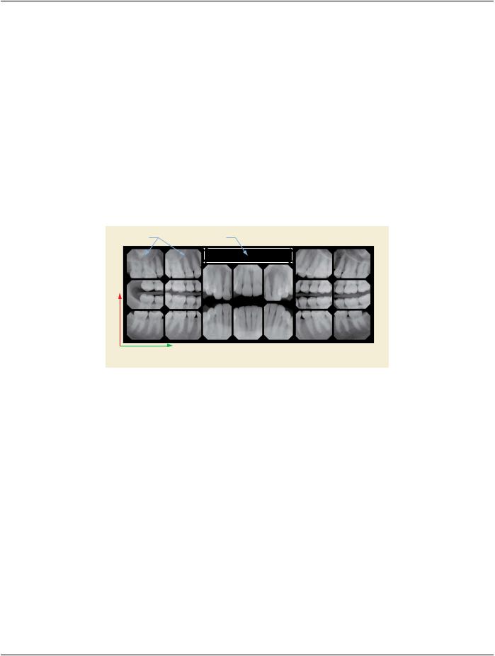
1.​A patient visits a General Dentist where a Full Mouth Series Exam with 18 images is acquired. The dentist observes severe bone​ loss and refers the patient to a Periodontist. The 18 images from the Full Mouth Series along with a Structured Display are copied​ to a DICOM Interchange CD and sent with the patient to see the specialist. The Periodontist uses the CD to open the exam in​ his Dental Radiographic Software and consults via phone with the General Dentist. Both are able to observe the same exam​ showing the images on each user's display using the exact same layout.​

Image Box

Text Box

(1.0, 1.0)


(0.0, 0.0)

(1.0, 0.0)

BLHC


Figure OO-1. Intra-oral Full Mouth Series Structured Display​

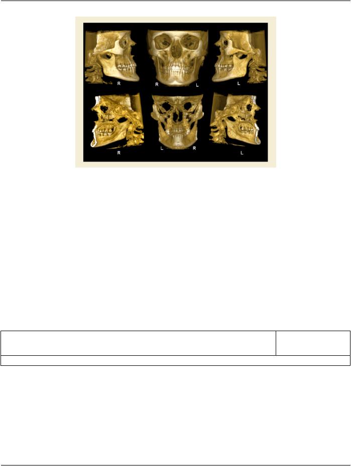
2.​A patient requests cosmetic surgery to enhance their facial appearance. The case requires consultation between an orthodontist​ in New York and an oral surgeon in California. The cephalometric series of 2D projections constructed from the volumetric CT​ data that is used for the discussion is arranged by a Structured Display for transfer between the two practitioners.​

- Standard -​

Page 358​

DICOM PS3.17 2020a - Explanatory Information​

Figure OO-2. Cephalometric Series Structured Display​

3.​AdentalproviderwishestocaptureaseriesofDICOMIOimagesforthepatient’sdentition.Thetoothmorphology,teetharedivided​ into molars, premolars, canines and incisors, and a number of images for each jaw. The anatomic information was captured​ utilizing the triplet of schema. This standard code sequence is based on ISO 3950-2010, Dentistry - Designation system for teeth​ and areas of the oral cavity.​

Every IO image should have anatomic information either through the primary or modifier sequence.​

In most standard cases, images are oriented in structured layouts. These structured displays are useful to be shared between​ providers for reference purposes.​

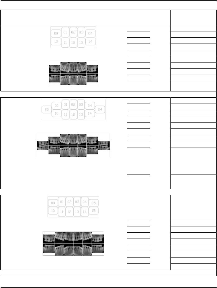
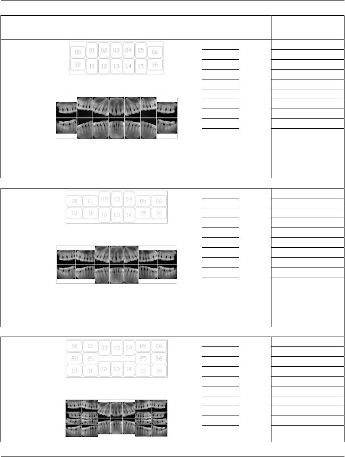
Table OO.1.1-1 shows structured display standard templates, where Viewset ID is based on the Japanese Society for Oral and​ Maxillofacial Radiology (JSOMR) classification provided by JIRA (Japan Medical Imaging and Radiological Systems Industries​ Association, www.jira-net.or.jp). Expected or typical teeth to be imaged location, region and designation codes are based on ISO​ 3950-2010, Dentistry - Designation system for teeth and areas of the oral cavity. For all the hanging protocols listed in OO.1.1-​ 1, the value to use for Hanging Protocol Creator (0072,0008) is "JSOMR" and the value to use for Hanging Protocol Name​ (0072,0002) does not include "JSOMR" (e.g., "DL-S001A", not "JSOMR DL-S001A").​

Table OO.1.1-1. Hanging Protocol Names for Dental Image Layout based on JSOMR classification​

Hanging​

Image​

ImageSize​ISO Teeth Designation​

Protocol​

Location​

(typical)​

Name​

Code​


10 Standard A Dental Image Layout​


- Standard -​

DICOM PS3.17 2020a - Explanatory Information​

Page 359​

Hanging​

Protocol​

Name​

DL-S001A​

Reference:​

10 Standard+2 Bitewing A Dental Image Layout​

DL-S002A​

Layout A Reference:​

10 Standard A Dental Image Layout​

DL-S003A​

Reference:​

Standard A Dental Image Layout​

Image​

ImageSize​ISO Teeth Designation​

Location​

(typical)​

Code​


00​

18, 17, 16, 15​

01​

14, 13, 12​

02​

12, 11, 21, 22​

03​

22, 23, 24​

04​

25, 26, 27, 28​

10​

48, 47, 46, 45​

11​

44, 43, 42​

12​

42, 41, 31, 32​

13​

32, 33, 34​

14​

35, 36, 37, 38​

00​

18, 17, 16, 15​

01​

14, 13, 12​

02​

12, 11, 21, 22​

03​

22, 23, 24​

04​

25, 26, 27​

10​

47, 46, 45​

11​

44, 43, 42​

12​

42, 41, 31, 32​

13​

32, 33, 34​

14​

35, 36, 37​

20​

18, 17, 16, 15,​


48, 47, 46, 45​

24​

25, 26, 27,28,​


35, 36, 37, 38​

00​

18, 17, 16, 15​

01​

14, 13, 12​

02​

12, 11​

03​

21, 22​

04​

22, 23, 24​

05​

25, 26, 27, 28​

10​

48, 47, 46, 45​

11​

44, 43, 42​

12​

42, 41​

13​

31, 32​

14​

32, 33, 34​

15​

35, 36, 37, 38​

- Standard -​

Page 360​

DICOM PS3.17 2020a - Explanatory Information​

Hanging​

Protocol​

Name​

DL-S004A​

Reference:​

14 Standard B Dental Image Layout​

DL-S004B​

Reference:​

14 Standard+4 Bitewing A Dental Image Layout​

DL-S005A​

Reference:​

Image​

ImageSize​ISO Teeth Designation​

Location​

(typical)​

Code​


00​

18, 17, 16​

01​

16, 15, 14​

02​

14, 13, 12​

03​

12, 11, 21, 22​

04​

22, 23, 24​

05​

24, 25, 26​

06​

26, 27, 28​

10​

48, 47, 46​

11​

46, 45, 44​

12​

44, 43, 42​

13​

42, 41, 31, 32​

14​

32, 33, 34​

15​

34, 35, 36​

16​

36, 37, 38​

00​

18, 17, 16​

01​

16, 15, 14​

02​

14, 13, 12​

03​

12, 11, 21, 22​

04​

22, 23, 24, 25​

05​

24, 25, 26, 27​

06​

25, 26, 27, 28​

10​

48, 47, 46, 45​

11​

47, 46, 45, 44​

12​

44, 43, 42​

13​

42, 41, 31, 32​

14​

32, 33, 34​

15​

34, 35, 36, 37​

16​

35, 36, 37, 38​

00​

18, 17, 16, 15​

01​

17, 16, 15, 14​

02​

14, 13, 12, 11,​

03​

12, 11, 21, 22​

04​

21, 22, 23, 24,​

05​

24, 25, 26​

06​

25, 26, 27, 28​

10​

48, 47, 46, 45​

11​

46, 45, 44, 43​

12​

44, 43, 42​

- Standard -​

Источник: https://studfile.net/preview/14585770/