Материал: Oxford American Handbook of Urology ( PDFDrive )

Внимание! Если размещение файла нарушает Ваши авторские права, то обязательно сообщите нам

662 CHAPTER 17 Basic science in urological practice

Other factors that influence RBF

•Sympathetic nerves innervate the glomerular arterioles. A reduction in circulating volume (such as blood loss) can stimulate sympathetic

nerves, causing the release of NA (which acts on 1-adrenoceptors on the afferent arteriole) to cause vasoconstriction. This results in reduced RBF and GFR (glomerular filtration rate).

•Angiotensin II constricts efferent arterioles and afferent arterioles and reduces RBF.

•Antidiuretic hormone (ADH), ATP, and endothelin all cause vasoconstriction and reduce RBF and GFR.

•Nitric oxide causes vasorelaxation and increases RBF.

•Atrial natriuretic peptide (ANP) causes afferent arteriole dilatation and increases RBF and GFR.

Renal function

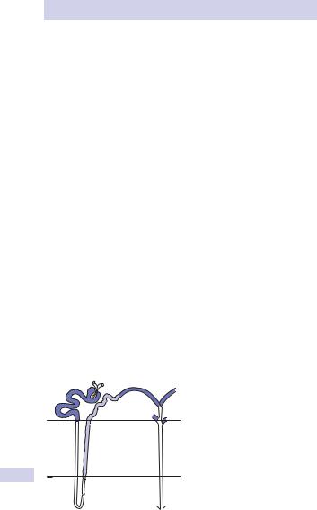
Each kidney has 1 million functional units, or nephrons (Fig. 17.1), the functional unit of the kidney consisting of a glomerular capillary network, surrounded by podocytes (epithelial cells) of Bowman’s capsule, which drains into a tubular system (proximal convoluted tubule, loop of Henle, distal convoluted tubule, collecting tube, and collecting duct) (Fig. 17.1).

Blood is delivered to the glomerular capillaries by an afferent arteriole and drained by an efferent arteriole. An ultrafiltrate of plasma is formed within the lumen of Bowman’s capsule, driven by Starling forces across the glomerular capillaries. Reabsorption of salt and water occurs in the proximal tubule, loop of Henle, distal tubule, and collecting ducts.

Clearance is the volume of plasma that is completely cleared of solute by the kidney per minute.

Glomerular filtration rate (GFR) is the clearance for any substance that is freely filtered and is neither reabsorbed, secreted, nor metabolized by the kidney.

Cortex

1


2


3


8

KEY:


7


1

Afferent and efferent arterioles


of the glomerulus

Outer


2

Bowman’s capsule

6


3

Proximal convoluted tubule

medulla

9

4

Loop of Henle (thin descending limb)

4


5

Loop of Henle (thin ascending limb)


6

Loop of Henle (thick ascending limb)


7

Distal convoluted tubule

Inner


8

Collecting tubule


9

Collecting duct

medulla

5


Figure 17.1 The nephron.

RENAL ANATOMY: RENAL BLOOD FLOW AND RENAL FUNCTION 663

For a substance that is freely filtered at the glomerulus, is neither secreted nor reabsorbed by the renal tubules, and is not metabolized (catabolized), clearance is equivalent to GFR. When a substance is both filtered at the glomerulus and secreted by the renal tubules, its clearance will be greater than GFR. When a substance is filtered at the glomerulus, but reabsorbed by the renal tubules, its clearance will be less than GFR.

GFR is directly related to RBF.

Experimentally, GFR can be accurately measured by measuring the clearance of inulin (a substance that is freely filtered by the glomerulus and is neither secreted nor reabsorbed by the kidneys). Thus, the volume of plasma from which in 1 minute the kidneys remove all inulin is equivalent to GFR. Normally about one-fifth (120 mL/min) of the plasma that flows through the glomerular capillaries (600 mL/min) is filtered.

Clinically, GFR is estimated using serum and urine creatinine, and is ~80–125 mL/min in an adult male and 75–115 mL/min in an adult female.

Clearance of a substance from the plasma can be expressed mathematically as

Clearance = U xV / P

where U is the concentration of a given substance in urine, P is its concentration in plasma, and V is the urine flow.

Clinically, a serum and 24-hour urine can be obtained to measure creatine clearance by the formula:

(Urine creatinine × total urine volume)

Clearance =

(plasma creatinine × time)

where time = 1440 minutes for a 24-hour urine collection

Alternatively, the eGFR (estimated GFR) is based on serum creatinine combined with other factors such as age, sex, and race is used more commonly than the 24-hour urine collection. Various on-line calculators are available for this eGFR calculation, such as http://www.nkdep.nih.gov/professionals/gfr_calculators/idms_con.htm.

The Modification of Diet in Renal Disease (MDRD) eGFR equation does not require weight as results are normalized to 1.732 body surface area (BSA), an accepted “average” adult BSA.1 The National Kidney Disease Education Program, sponsored by the NIH, recommends reporting estimated GFR values greater than or equal to 60 mL/min/1.73 m2 simply as “t60 mL/min/1.73 m2,” not an exact number.

eGFR (mL/min/1.73 m2) = 186 x (Scr)-1.154 x (Age)-0.203

x (0.742 if female) x (1.212 if African American) (conventional units)

1 Levey AS, Bosch JP, Lewis JB, et al. (1999). A more accurate method to estimate glomerular filtration rate from serum creatinine: a new prediction equation. Modification of Diet in Renal Disease Study Group. Ann Intern Med 130(6):461–470.

664 CHAPTER 17 Basic science in urological practice

Renal physiology: regulation of water balance

Total body water (TBW) is 42 L. It is contained in two major com- partments—the intracellular fluid (ICF or the water inside cells) which accounts for 28 L and the extracellular fluid (ECF or water outside cells) representing 14 L.

ECF is further divided into interstitial fluid (ISF, 11 L), transcellular fluid (1 L), and plasma (3 L). Hydrostatic and osmotic pressures influence movement between the compartments. Water is taken in from fluids, food, and oxidation of food.

Water is lost from urine, feces, and insensible losses. Intake and losses usually balance (~2 L/day) and TBW remains relatively constant.

Antidiuretic hormone (ADH or vasopressin)

ADH is secreted from the posterior pituitary in response to stimuli from changes in plasma osmolarity (detected by osmoreceptors in the hypothalamus) or changes in blood pressure or volume (detected by baroreceptors in the left atrium, aortic arch, and carotid sinus). These changes also stimulate the thirst center in the brain.

The action of ADH on the kidney

•Increases collecting duct permeability to water and urea

•Increases loop of Henle and collecting duct reabsorption of NaCl

•Increases vasoconstriction

Conditions of water excess

Water excess occurs when body fluids become hypotonic and ADH release and thirst are suppressed. In the absence of ADH, the collecting duct is impermeable to water and a large volume of hypotonic urine is produced, thus restoring normal plasma osmolarity.

Conditions of water deficit

When body fluids are hypertonic, ADH secretion and thirst are stimulated. The collecting duct becomes permeable, water is reabsorbed into the lumen, and a small volume of hypertonic urine is excreted.

The ability to concentrate or dilute urine depends on the countercurrent multiplication system in the loop of Henle. Essentially, a medullary concentration gradient is generated (partly by the active transport of NaCl), which provides the osmotic driving force for the reabsorption of water from the lumen of the collecting duct when ADH is present.

Children have a circadian rhythm in ADH secretion—high at night and low during the day. Adults essentially have a constant ADH secretion over a 24-hour period, with slight increases occurring around mealtimes. At these times, increased ADH secretion probably acts to prevent sudden increases in plasma osmolality that would otherwise occur due to ingestion of solutes in a meal.

RENAL PHYSIOLOGY: REGULATION OF NA AND K EXCRETION 665

Renal physiology: regulation of sodium and potassium excretion

Sodium regulation

NaCl is the main determinant of ECF osmolality and volume. Osmolality = osmoles per kg water (osmol/kg). Osmolarity = osmoles per liter of solution (osmol/L.

Low-pressure receptors in the pulmonary vasculature and cardiac atria, and high-pressure baroreceptors in the aortic arch and carotid sinus recognize changes in the circulating volume. Decreased blood volume triggers increased sympathetic nerve activity and stimulates ADH secretion, which results in reduced NaCl excretion.

Conversely, when blood volumes are increased, sympathetic activity and ADH secretion are suppressed, and NaCl excretion is enhanced (natriuresis).

A variety of natriuretic peptides have been isolated that cause a natriuresis. Under physiological conditions, renal natriuretic peptide (urodilatin) is the most important of these. Atrial natriuretic hormone (ANP) may influence sodium output under conditions of heart failure.

Renin–angiotensin–aldosterone system

Renin is an enzyme made and stored in the juxtaglomerular cells found in the walls of the afferent arteriole. Factors increasing renin secretion are as follows:

•Reduced perfusion of the afferent arteriole

•Sympathetic nerve activity

•Reduced Na+ delivery to the macula densa

Renin acts on angiotensin to create angiotensin I. This is converted to angiotensin II in the lungs by angiotensin-converting enzyme (ACE). Angiotensin II performs several functions, which result in the retention of salt and water:

•Stimulates aldosterone secretion (resulting in NaCl reabsorption)

•Vasoconstriction of arterioles

•Stimulates ADH secretion and thirst

•Enhances NaCl reabsorption by the proximal tubule

Potassium regulation

K+ is critical for many cell functions. A large concentration gradient across cell membranes is maintained by the Na+-K+-ATPase pump. Insulin and adrenaline also promotes cellular uptake of K+.

The kidney excretes up to 95% of K+ ingested in the diet. The distal tubule and collecting duct are able to both reabsorb and secrete K+. Factors promoting K+ secretion include the following:

•Increased dietary K+ (driven by the electrochemical gradient)

•Aldosterone

•Increased rate of flow of tubular fluid (i.e., diuretics)

•Metabolic alkalosis (acidosis exerts the opposite effect)

666 CHAPTER 17 Basic science in urological practice

Renal physiology: acid–base balance

The normal pH of extracellular fluid (ECF) is 7.4 ([H+] = 40 nmol/L). Several mechanisms are in place to eliminate acid produced by the body and maintain body pH within a narrow range.

Buffering systems limiting [H+] fluctuation in the blood

Buffer bases that take up H+ ions in the body include the following:1

Bicarbonate buffer system

H+ + HCO3– l H2CO3 l H2O + CO2

Phosphate system

H+ + HPO4–2 l H2PO4–

Protein buffers

H+ + Protein– l HProtein

The Henderson–Hasselbalch equation describes the relationship between pH and the concentration of conjugate acid and base:

From this equation, it can be seen that alterations in bicarbonate [HCO3–] or CO2 will affect pH. Metabolic acid–base disturbances relate to a change in bicarbonate, and respiratory acid–base disorders relate to alterations in CO2.

Bicarbonate reabsorption along the nephron

Bicarbonate is the main buffer of ECF and is regulated by both the kidneys and lungs; 85% is reabsorbed in the proximal convoluted tubule (Fig. 17.2).

Carbonic acid is first produced from CO2 and water (accelerated by carbonic anhydrase). The carbonic acid dissociates, and an active ion pump (Na+/H+ antiporter) extrudes intracellular H+ into the tubule lumen in exchange for Na+. Secretion of H+ ions favors a shift of the carbonic acid– bicarbonate equilibrium toward carbonic acid, which is rapidly converted into carbon dioxide and water.

CO2 diffuses into the tubular cells down its diffusion gradient and is reformed into carbonic acid by intracellular carbonic anhydrase. The bicarbonate formed by this reaction is exchanged for chloride and passes into the circulation. Essentially, with each H+ ion that enters the kidney, a bicarbonate ion enters the blood, which bolsters the buffering capacity of the ECF.

The remaining bicarbonate is absorbed in the distal convoluted tubule, where cells actively secrete H+ into the lumen via an ATP-dependent pump. The distal tubule is the main site that pumps H+ into the urine to ensure the complete removal of bicarbonate. Once the bicarbonate has gone, phosphate ions and ammonia buffer any remaining H+ ions.

1 H2O = water; CO2 = carbon dioxide; HCO3– = bicarbonate; H2CO3 = carbonic acid; H+ = hydrogen ions; H2PO42– = dihydrogen phosphate; H3PO4 = phosphoric acid.

Источник: https://studfile.net/preview/16690383/