Материал: Oxford American Handbook of Urology ( PDFDrive )

42 CHAPTER 2 Urological investigations
Uses of plain abdominal radiography (KUB X-ray—kidneys, ureters, bladder)
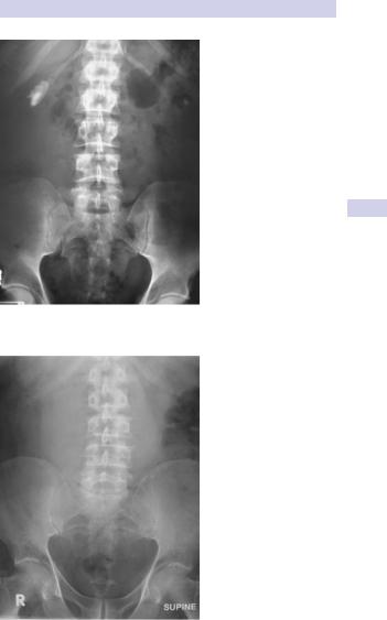
Plain abdominal radiography is for detection of stones and determination of their size and (to an extent) their location within the kidneys, ureters, and bladder (Fig. 2.3).
For renal calculi, a calcification overlying the kidneys is intrarenal if it maintains its relationship to the kidney on inspiratory and expiratory films (i.e., if it moves with the kidney). If in doubt as to whether an opacity overlying the outline of the kidney is intrarenal or not, get an ultrasound (look for the characteristic acoustic shadow within the kidney), intravenous pyelography (IVP), or computed tomographic urography (CTU).
Sensitivity for detection of renal calculi is on the order of 50–70% (i.e., the false-negative rate is between 30% and 50%; it misses ureteric stones when these are present in 30–50% of cases). CTU or IVP, which relate the position of the opacity to the anatomical location of the ureters, are required to make a definitive diagnosis of a ureteric stone. However, once the presence of a ureteric stone has been confirmed by another imaging study (CTU or IVP), and as long as it is radio-opaque enough and large enough to be seen, plain radiography is a good way of following the patient to establish whether the stone is progressing distally, down the ureter.
Plain radiography is not useful for following ureteric stones that are radiolucent (e.g., uric acid) or small (generally a stone must be 3–4 mm to be visible on plain X-ray), or when the stones pass through the ureter as it lies over the sacrum. The ability of KUB X-ray to visualize stones is also dependent on the amount of overlying bowel gas.
Plain tomography (a plain X-ray taken of a fixed coronal plane through the kidneys) can be useful, but is rarely done with the availability of ultrasound and CT.
Opacities that may be confused with stones (renal, ureteric) on plain radiography include calcified lymph nodes and pelvic phleboliths (round, lucent center, usually below the ischial spines).
Look for the psoas shadow—this is obscured where there is retroperitoneal fluid (pus or blood) (Fig. 2.4).
USES OF PLAIN ABDOMINAL RADIOGRAPHY 43
Figure 2.3 Small staghorn calculus on KUB X-ray.
Figure 2.4 Leaking abdominal aortic aneurysm (AAA) on plain X-ray; the right psoas shadow cannot be seen because of retroperitoneal hemorrhage.

44 CHAPTER 2 Urological investigations
Intravenous pyelography (IVP)
IVP is also known as intravenous urography (IVU). A control film is obtained before contrast is given. Intravascular contrast is administered followed by a series of X-rays of the kidneys, ureters, and bladder over the following 30 minutes or so, to image their anatomy and pathology, and to give some indication of renal function.
•Radio-opacity of contrast agents depends on the presence of a triiodinated benzene ring in the molecule.
•Ionic monomers (sodium and meglumine salts) ionize, thereby producing high-osmolality solutions (e.g., iothalamate—Conray), diatrizoate—Hypaque, Urografin).
•Nonionic monomers have low osmolality (e.g., iopamidol—Niopam, iohexol—Omnipaque).
•At a concentration of 300 mg of iodine per mL, ionic monomers have an osmolality 5xhigher than that of plasma, compared with nonionic monomers, which have an osmolality 2xthat of plasma.
•Excreted from plasma by glomerular filtration.
Films and “phases” of IVP
Plain film
This is used to look for calcification overlying the region of the kidneys, ureters, and bladder.
Nephrogram phase
This is the first phase of IVP; film is taken immediately following intravenous administration of contrast (peak nephrogram density). The nephrogram is produced by filtered contrast within the lumen of the proximal convoluted tubule (it is a proximal tubular, rather than distal tubular, phenomenon).
Pyelogram phase
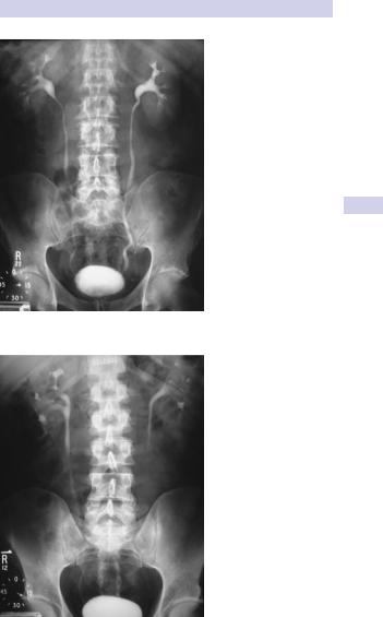
As the contrast passes along the renal tubule (into the distal tubule) it is concentrated (as water is absorbed, but the contrast agent is not). As a consequence, the contrast medium is concentrated in the pelvicalyceal system, thus this pyelogram phase (Fig. 2.5) is much denser than the nephrogram phase.
The pyelogram phase can be made denser by dehydrating the patient prior to contrast administration. Pelvic compression can be used to distend the pelvicalyceal system and demonstrate their anatomy more precisely. Compression is released and a film taken (20–30 min) (Fig. 2.6).
Side effects of administration of intravenous contrast media
These occur in 1% of patients given nonionic and 5% given ionic contrast media.
The most serious reactions represent an anaphylactic reaction—hypo- tension with flushing of the skin (marked peripheral vasodilatation), edema (face, neck, body, and limbs), bronchospasm, and urticaria. Rarely, cardiac arrest can occur. The death rate, as a consequence of these reactions, is ~1 in 40,000 to 1 in 70,000 with the ionic media, and ~1 in 200,000 with nonionic contrast agents.
INTRAVENOUS PYELOGRAPHY (IVP) 45
Figure 2.5 Normal IVP at 15 minutes.
Figure 2.6 Normal IVP at 20 minutes. Lower abdominal compression has been released.

46 CHAPTER 2 Urological investigations
A contrast reaction is more likely to occur in patients with an iodine allergy, previous contrast reaction, asthma, multiple other allergies, and heart disease and is less likely with nonionic contrast media. Steroid premedication (prednisone 50 mg given at 13, 7, and 1 hour prior to the procedure) can reduce the risk of a contrast reaction.
Contrast media are also nephrotoxic; 10% of patients with a raised creatinine will develop an increase in creatinine after an IVP (more likely in diabetics, with dehydration, and with large contrast doses). The increase in creatinine usually resolves spontaneously.
Uses of IVP
•Investigation of hematuria—detection of renal masses, filling defects within the collecting system of the kidney and within the ureters (stones, TCCs)
•Localization of calcification overlying the urinary tract (i.e., is it a stone or not?)
•Investigation of patients with flank pain (e.g., suspected ureteric colic). IVP is increasingly being replaced with CTU, which has superior sensitivity and specificity.
•Very good for identification of congenital urinary tract abnormalities (e.g., ureteric anatomy in duplex systems) (Fig. 2.7); malrotation; horseshoe kidneys
•Used for follow-up postureteric surgery to identify strictures