Материал: Oxford American Handbook of Urology ( PDFDrive )

TESTICULAR CANCER: CLINICAL PRESENTATION 297
Box 6.6 Differential diagnosis of testicular and scrotal masses in adults and children
Adult or pediatric painful mass
•Epididymitis/orchitis; bacterial, STD, mumps, tuberculosis
•Incarcerated/strangulated hernia
•Testicular trauma: usually blunt; contusion, rupture; usually associated hematocele
•Torsion (testicle, testicular or epididymal appendage)
•Tumor (pain infrequent unless traumatized or rapidly growing; see below)
Adult painless mass
•Adenomatoid tumor of testis or epididymis
•Adrenal rest tumors
•Adenocarcinoma of the rete testis
•Chylocele: usually associated with filariasis
•Fibrous pseudotumor of the tunica albuginea
•Hydrocele, primary or due to trauma, torsion, tumor, epididymitis; hydrocele of the cord
•Lipoma of the cord
•Mesothelioma of tunica vaginalis
•Polyorchidism
•Paratesticular sarcomas: rhabdomyosarcoma, fibrosarcoma, leiomyosarcoma, liposarcoma
•Scrotal edema (insect bite, nephrotic syndrome, acute idiopathic scrotal edema)
•Scrotal wall: sebaceous and inclusion cysts, idiopathic calcinosis, fat necrosis, malignancy
•Sperm granuloma following vasectomy
•Spermatocele (epididymal cyst)
•Testicular cysts (simple, tunica albuginea, epidermoid)
•Testicular tumor
•Germ cell tumors (95% of testicular malignancies): seminoma, embryonal cell carcinoma, choriocarcinoma, yolk sac carcinoma teratoma (1–5%), teratocarcinoma
•Gonadal stromal tumors: Leydig, Sertoli cell, granulosa cell tumor
•Metastatic tumors: prostate, lung, and gastrointestinal tract; rare kidney, malignant melanoma, pancreas, bladder and thyroid
•Mixed germ cell and stromal tumor (gonadoblastoma)
•Angioma, fibroma, leiomyoma, hamartoma, carcinoid, mesothelioma, and neurofibroma
•Malignant fibrous histiocytoma (most common soft tissue sarcoma in late adult life)
•Leukemia or lymphoma
•Varicocele
Pediatric painless mass
•Similar to adult list; most/more common: hydrocele, hernia, variocele, testicular teratoma, adrenal rest tumor, rhabdomyosarcoma
Reproduced from Carver BS, Sheinfeld J (2010). Testis cancer, general. Gomella LG (Ed.), 5 Minute Urology Consult, 2nd ed. Philadelphia LWW.

298 CHAPTER 6 Urological neoplasia
Ultrasound may also be used to identify impalpable lesions as small as 1–2 mm—an occult primary tumor in a patient presenting with systemic symptoms and signs or an incidental finding.
Image contralateral testis as 2% of patients will have bilateral testicular cancers.
Abdominal and chest CT scans are usually obtained for staging purposes if the diagnosis of TC is confirmed, usually following radical orchiectomy.
Serum tumor markers are measured prior to any treatment of a confirmed testicular mass (p. 298).
Treatment
All patients with a testicular mass should be evaluated for testicular cancer. Studies have shown that 18–33% of patients with testicular cancer were initially treated for epididymitis, resulting in a delay in diagnosis.
Radical orchiectomy
Radical orchiectomy should be performed for diagnosis and treatment of the primary tumor. This involves excision of the testis, epididymis, and cord, with their coverings, through a groin incision. The cord is clamped, transfixed, and divided near the internal inguinal ring before the testis is manipulated into the wound, preventing inadvertent metastasis.
A silicone prosthesis may be inserted at the time of the procedure or at a later date. This treatment is curative in up to 80% of patients. Sperm cryopreservation should be offered to patients without a normal contralateral testis.
Contralateral testis biopsy should be considered in patients at high risk for IGCN (see pp. 294, 308).

TESTICULAR CANCER: SERUM MARKERS 299
Testicular cancer: serum markers
Germ cell tumors may express and secrete into the bloodstream relatively specific and readily measurable proteins. These tumor markers (with the exception of PLAP) are useful in diagnosis, staging, prognostication (see
p.303), and monitoring of response to treatment (see p. 304).
Currently, testicular cancer is the only malignancy to incorporate serum
markers into the staging system.
Oncofetal proteins
A-Fetoprotein (AFP)
AFP is expressed by trophoblastic elements within 50–70% of teratomas and yolk sac tumors. With respect to seminoma, the presence of elevated serum AFP strongly suggests a non-seminomatous element.
Serum half-life is 3–5 days; normal is <10 ng/mL. It can be elevated.
Human chorionic gonadotrophin, B subunit (B-hCG)
B-hCG is expressed syncytiotrophoblastic elements of choriocarcinomas (100%), teratomas (40%), and seminomas (10%). Serum half-life is 24–36 hours. Assays measure the Bsubunit with the normal <5 mIU/mL.
When used together, 90% of patients with advanced disease have elevation of one or both markers; it is less among patients with low-stage tumors.
Cellular enzymes
Lactate dehydrogenase (LDH)
LDH is a ubiquitous enzyme, elevated in serum for various causes and is therefore less specific. It is elevated in 10–20% of seminomas, correlating with tumor burden, and is most useful in monitoring treatment response in advanced seminoma.
Other markers with limited current clinical use include PLAP, CD30, and GGTP.
Clinical use
These markers are measured at presentation, 1–2 weeks after radical orchiectomy, and during follow-up to assess response to treatment and residual disease.
Normal markers prior to orchiectomy do not exclude metastatic disease. Normalization of markers post-orchiectomy cannot be equated with absence of disease, and persistent elevations of markers postorchiectomy may occur with liver dysfunction and hypogonadotrophism, but usually indicate metastatic disease.

300 CHAPTER 6 Urological neoplasia
Testicular cancer: pathology and staging
Ninety percent of testicular tumors are malignant germ cell tumors (GCT), split into seminomatous and non-seminomatous (NS) GCTs for clinical purposes. Seminoma, the most common germ cell tumor, appears pale and homogeneous. NSGCTs are heterogeneous and sometimes contain bizarre tissues such as cartilage or hair.
Metastases to the testis are rare, notably from the prostate (35%), lung (19%), colon (9%), and kidney (7%). Table 6.15 shows the WHO histopathological classification of testicular tumors.
The right testis is affected slightly more commonly than the left; synchronous bilateral TC occurs in 2% of cases. TC spreads by local extension into the epididymis, spermatic cord, and, rarely, the scrotal wall.
Lymphatic spread occurs via the testicular vessels, initially to the paraaortic nodes. Involvement of the epididymis, spermatic cord, or scrotum may lead to pelvic and inguinal node metastasis.
Blood-borne metastasis to the lungs, liver, and bones is more likely once the disease has breached the tunica albuginea.
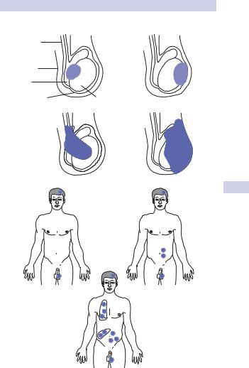
TC is staged using various classifications, most recently the TNM (2002) system (see Fig. 6.9 and Table 6.16). Here, T stage is pathological, N stage involves imaging, and M stage involves physical examination, imaging, and biochemical investigations. An additional S category is appended for serum tumor markers (see p. 305).
Table 6.15 WHO histopathological classification of testicular tumors
Germ cell tumors (90%)
Seminoma (48%)
•Spermatocytic, classical, and anaplastic subtypes
Non-seminomatous GCT (42%)
•Teratoma
•Differentiated/mature
•Intermediate/immature
•Undifferentiated/malignant
•Yolk sac tumor
•Choriocarcinoma
•Mixed NSGCT
Mixed GCT (10%)
Other tumors (7%)
•Epidermoid cyst (benign)
•Adenomatoid tumor
•Adenocarcinoma of the rete testis
•Carcinoid
•Lymphoma (5%)
•Metastatic, from another site (1%)
Sex cord stromal tumors (3%) (10% malignant)
•Leydig cell
•Sertoli cell
•Mixed or unclassified
Mixed germ cell/sex cord tumors (rare)
TESTICULAR CANCER: PATHOLOGY AND STAGING 301
(a)
Vas deferens, spermatic cord
Scrotal wall
Epididymis
Tunica vaginalis
(b)
T1−4
T1 |
T2 |
Testis
T3 |
T4 |
N0 M0 |
T1−4 N1−3 M0 |
T1−4 N1−3 M1
Figure 6.9 Pathological staging of testicular cancer. (a) Primary tumors T1–T4. If radical orchiectomy has not been used, Tx is used. (b) Node/metastasis: para-aortic lymphadenopathy, measured in long axis on CT scan (upper figures); supraclavicular lymphadenopathy and/or pulmonary metastases—M1a, other distant metastases (e.g., liver, brain)—M1b (lower figure).