Материал: Oxford American Handbook of Urology ( PDFDrive )

312 |
CHAPTER 6 Urological neoplasia |
|
|
|
|
Table 6.19 Continued |
|
|
|
|
|
|
|
|
|
Disease (organism/ |
Characteristics |
Therapy |
|
|
cause) |
|
|
|
|
|
|
|
|
|
Lymphogranuloma |
Small, painless vesicle or |
Doxycycline; |
|
|
venereum (Chlamydia |
papule progresses to an |
azithromycin or |
|
|
trachomatis) |
ulcer |
erythromycin |
|
|
|
Painful matted nodes, fistula |
alternative |
|
|
|
common |
|
|
|
Molluscum contagiosum |
Smooth, round, pearly |
Surgical ablation |
|
|
(A pox virus) |
papules, 2–5 mm |
|
|
|
Pearly penile papules |
Usually uncircumcised, |
Usually not clinically |
|
|
(? viral) |
painless 1–2 mm papules |
important; ablation if |
|
|
|
with variable color |
bothersome |
|
|
|
(translucent/white/yellow) |
|
|
|
|
usually around corona an |
|
|
|
|
dorsum of penis |
|
|
|
Sclerosing lymphangitis |
Asymptomatic |
|
(Local trauma) |
subcutaneous cordlike |
|
|
swellings along the dorsal |
|
|
shaft of the penis or around |
|
|
the coronal sulcus; may be |
|
|
edema of glans and coronal |
|
|
sulcus. |
|
Syphilis, primary |
Painless, indurated, with a |
|
(Treponema pallidum) |
clean base, usually singular |
|
|
lesion; tender inguinal |
|
|
nodes |
Traumatic in origin, selflimited; avoid vigorous sexual activity
Benzthiazide penicillin G (2.4 million units IM) single dose
Zoon balanitis (? |
Found in uncircumcised |
Circumcision curative |
Mycobacterium |
only, on prepuce or glans, |
|
smegmatis) |
usually painless, solitary |
|
|
shiny lesion with reddish or |
|
|
orange tint; can be to 2 cm, |
|
|
occasionally erosive |
|
|
|
|
•Cutaneous horn: rare, solid skin overgrowth; extreme hyperkeratosis, the base may be malignant. Treatment is wide local excision.
•Pseudoepitheliomatous micaceous and keratotic balanitis: unusual hyperkeratotic growths on the glans. They require excision, histological examination, and follow-up, as they may recur.
•Balanitis xerotica obliterans (BXO): also known as lichen sclerosus et atrophicus, this is a common sclerosing condition of glans and prepuce. It occurs at all ages and most commonly presents as non-retractile foreskin (phimosis). The meatus and fossa navicularis may be affected, causing obstructed and spraying voiding. The histological diagnosis is usually made after circumcision, with epithelial atrophy, loss of rete pegs, and collagenization of the dermis. BXO occurs in association

PENILE NEOPLASIA 313
with penile SCC, but most pathologists would regard the lesion as benign unless epithelial dysplasia is present.
•Leukoplakia: solitary or multiple whitish glanular plaques that usually involve the meatus. Treatment is excision and histology. Leukoplakia is associated with in situ SCC; follow-up is required.
•Erythroplasia of Queyrat: also known as carcinoma in situ of the glans, prepuce, or penile shaft. A red, velvety, circumscribed painless lesion occurs, though it may ulcerate, resulting in discharge and pain. Treatment is excision biopsy if possible; radiotherapy, laser ablation, or topical 5-fluorouracil may be required. Histology reveals hyperplastic mucosal cells with malignant features.
•Bowen disease: this is carcinoma in situ of the remainder of the keratinizing genital or perineal skin. Treatment is wide local excision, laser, or cryoablation.
•Buschke–Löwenstein tumor: also known as verrucous carcinoma or giant condyloma acuminatum, this is an aggressive locally invasive
tumor of the glans. Metastasis is rare, but wide excision is necessary to distinguish it from SCC. Urethral erosion and fistulation may occur.

314 CHAPTER 6 Urological neoplasia
Penile cancer: epidemiology, risk factors, and pathology
Squamous cell carcinoma (SCC) is the most common penile cancer, accounting for 95% of penile malignancies. Others include Kaposi sarcoma (see p. 310) and, rarely, basal cell carcinoma, melanoma, sarcoma, and Paget disease. Metastases to the penis are occasionally seen from the bladder, prostate, rectum, and other primary sites.
Incidence and etiology of SCC
Penile cancer is rare, representing 1% of male cancers. The incidence appears to be decreasing, occurring mostly in elderly men. In 2009, 1290 cases were reported, with 300 deaths.
Risk factors for SCC
•Age: penile cancer incidence rises during the sixth decade and peaks in the eighth decade. It is unusual <40 years of age, but has been rarely reported in children.
•Premalignant lesions: 42% of patients with penile SCC are reported to have had a pre-existing penile lesion (see p. 310).
•A prepuce (foreskin): penile cancer is rare in men circumcised at a young age. Smegma that forms from desquamated epithelial cells is thought to be a primary instigating factor in penile cancer; good hygiene and circumcision limit smegma accumulation.
•Geography: Highest incidence worldwide is in Brazil. It is virtually non-existent in Israel.
•Human papilloma virus (HPV) genital wart infection, especially with types 16, 18, and 21.
•Multiple sex partners
•Smoking and tobacco products
Pathology and staging of penile SCC
Believed to be preceded by carcinoma in situ, SCC starts as a slow-grow- ing papillary, flat or ulcerative lesion on the glans (48%), prepuce (21%), glans and prepuce (9%), coronal sulcus (6%), or shaft (2%). The remainder are indeterminate. It grows locally beneath the foreskin before invading the corpora cavernosa, urethra, and, eventually, the perineum, pelvis, and prostate.
Metastasis is initially to the superficial then deep inguinal and, subsequently, iliac and obturator lymph nodes. Skin necrosis, ulceration, and infection of the inguinal lymph nodes may lead to sepsis or hemorrhage from the femoral vessels. Blood-borne metastasis to lungs and liver is rare (1–10% of cases).
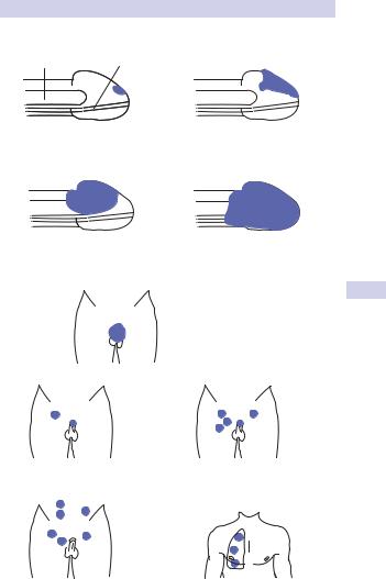
Histologically, SCC exhibits keratinization, epithelial pearl formation, and mitoses. Staging is by the TNM system (see Fig. 6.10 and Table 6.20). Grading scale is as follows: low (75%), intermediate (15%), or high (10%); grading correlates with prognosis, as does presence of vascular invasion.
PENILE CANCER: EPIDEMIOLOGY, RISK FACTORS, & PATHOLOGY 315
(a)Carpus
cavernosum Urethra
T1s or Ta |
T1 |
Epithelium only |
Subepithelia |
|
tissue invadded |
T2 |
T3 |
Corpus cavernosum |
Urethra/prostate |
or spongiosum invaded |
invaded |
|
T4 |
|
Scrotum, perineum |
|
bone invaded |
(b)
N1 – Single superficial |
N2 – multiple or bilateral |
ingunal node |
superficial ingunal |
|
nodes |
N3 – unilateral or |
M1 – distant metastasis |
bilateral ingunal |
(e.g. pulmonary) |
or intra-pelvic nodes |
|
Figure 6.10 Pathological staging of penile cancer. (a) T stage: primary tumor;
(b) N and M stages.

316 CHAPTER 6 Urological neoplasia
Table 6.20 TNM staging of penile carcinoma
|
|
Tx |
Primary tumor cannot be assessed |
|
|
T0 |
No evidence of primary tumor |
|
|
Tis |
Carcinoma in situ |
|
|
Ta |
Noninvasive verrucous carcinoma |
|
|
T1 |
Tumor invades subepithelial connective tissue |
|
|
T2 |
Tumor invades corpus cavernosum or spongiosum |
|
|
T3 |
Tumor invades urethra or prostate |
|
|
T4 |
Tumor invades other structures |
|
|
Nx |
Regional nodes cannot be assessed |
|
|
N0 |
No regional lymph node metastases |
|
|
N1 |
Single superficial inguinal lymph node metastasis |
|
|
N2 |
Multiple or bilateral superficial inguinal lymph node metastases |
|
|
N3 |
Metastases in deep inguinal or pelvic lymph nodes, unilateral or |
|
|
|
bilateral |
|
|
Mx |
Distant metastasis cannot be assessed |
|
|
M0 |
No distant metastasis |
|
|
M1 |
Distant metastasis |
|
|
|
|
SCCs are graded using the Broder classification system:
•Grade I: well differentiated, keratinization, prominent intercellular bridges, keratin pearls
•Grade II to III: greater nuclear atypia, increased mitotic activity, decreased keratin pearls
•Grade IV: cells deeply invasive, marked nuclear pleomorphism, nuclear mitoses, necrosis, lymphatic and perineural invasion, no keratin pearls