Материал: Oxford American Handbook of Urology ( PDFDrive )

342 CHAPTER 7 Miscellaneous urological diseases of kidney
Vesicoureteric reflux (VUR) in adults
VUR is the retrograde flow of urine from the bladder into the upper urinary tract with or without dilatation of the ureter, renal pelvis, and calyces. It can cause symptoms and may lead to renal failure (reflux nephropathy). In adults 10–15% of patients requiring hemodialysis or transplantation do so because of reflux nephropathy.
Pathophysiology
Reflux is normally prevented by low bladder pressures, efficient ureteric peristalsis, and the ability of the vesicoureteric junction (VUJ) to occlude the distal ureter during bladder contraction. This is assisted by the ureter passing obliquely through the bladder wall (the “intramural” ureter), which is 1–2 cm long. Normal intramural ureteric length to ureteric diameter ratio is 5:1.
VUR of childhood tends to resolve spontaneously with increasing age because as the bladder grows, the intramural ureter lengthens.
Classification
Primary
A primary anatomical (and therefore functional) defect is where the intramural length of the ureter is too short (ratio <5:1).
Secondary
A secondary defect is secondary to some other anatomical or functional problem:
-Bladder outlet obstruction (BPO, DSD due to neuropathic disorders,1 posterior urethral valves, urethral stricture) that leads to elevated bladder pressures
-Poor bladder compliance or the intermittently elevated pressures of detrusor hyperreflexia (due to neuropathic disorders1—e.g., spinal cord injury, spina bifida)
-Iatrogenic reflux following TURP or TURBT (a tumor overlying the ureteric orifice)—this is rare; ureteric meatotomy (incision of the ureteric orifice) for removal of ureteric stones at the VUJ; following incision of a ureterocele; ureteroneocystostomy; after pelvic radiotherapy
-Inflammatory conditions affecting function of the VUJ: TB, schistosomiasis, UTI
Associated disorders
VUR is commonly seen in duplex ureters (the Meyer–Weigert law).2 Cystitis can cause VUR through bladder inflammation, reduced bladder compliance, increased pressures, and distortion of the VUJ. Coexistence of UTI with VUR is a potent cause of pyelonephritis—reflux of infected urine under high pressure causes reflux nephropathy, resulting in renal scarring, hypertension, and renal impairment.
1 Neuropathic disorders therefore cause VUR because they lead to intermittently or chronically raised bladder pressure (due to BOO, poor compliance, and/or detrusor hyperreflexia).
2 The lower pole ureter inserts into the bladder in a proximal location to the upper pole ureter, which inserts distally—i.e., nearer the bladder neck. The lower pole ureter has a shorter intramural length and therefore refluxes. The upper pole ureter has a longer intramural length and tends to be obstructed.

VESICOURETERIC REFLUX (VUR) IN ADULTS 343
Presentation
-VUR may be symptomless, being identified during VCUG, IVP, or renal ultrasound (which shows ureteric and renal pelvis dilatation) done for some other cause.
-UTI symptoms
-Flank pain associated with a full bladder or immediately after micturition
Symptoms of recurrent UTI or of flank pain may have been present for many years before the patient seeks medical advice. Even then it may take some time for a diagnosis of VUR to be made because a high index of suspicion is required and the definitive test for making a diagnosis of VUR (VCUG—see below) is invasive (although VUR may be diagnosed by the less invasive use of IVP).
Investigation
The definitive test for the diagnosis of VUR is cystography. VUR may be apparent during bladder filling or during voiding (voiding cystourethrography [VCUG]—also known as micturating cystourethrography [MCUG]).
Urodynamics establishes the presence of voiding dysfunction (e.g., DSD) if this is suspected from the clinical picture. If there is radiographic evidence of reflux nephropathy, check blood pressure, check the urine for proteinuria, measure serum creatinine, and arrange for a 99mTc-DMSA isotope study to assess renal cortical scarring and determine split renal function.
Management
VUR is harmful to the kidney:
-In the presence of infected urine
-Where bladder pressures are markedly elevated (due to severe BOO, poor compliance, or high-pressure hyperreflexic bladder contractions)
In the absence of urinary infection or severe outflow obstruction/ raised bladder pressures, VUR is not harmful, at least in the short term (months).
Subsequent management depends on the following:
-The presence and severity of symptoms
-The presence of recurrent, bacteriologically proven urinary infection
-The presence of already established renal damage (radiological evidence of reflux nephropathy, hypertension, proteinuria— proteinuria is a poor prognostic factor in patients with VUR, indicating the likelihood of impending end-stage renal failure)
For the patient with primary VUR, recurrent UTIs with no symptoms between infections, no hypertension, and good renal function, treat the UTIs when they occur and consider low-dose antibiotic prophylaxis if UTIs occur frequently (say >3 per year). If the UTIs are regularly associated with constitutional disturbance (acute pyelonephritis rather than simple cystitis), then ureteric reimplantation is indicated.

344 CHAPTER 7 Miscellaneous urological diseases of kidney
For the patient with primary VUR and objective evidence of deterioration in the affected kidney (i.e., progressive radiological signs of reflux nephropathy or reduction in renal function), use ureteric reimplantation.
Reflux into a nonfunctioning kidney (<10% function on DMSA scan) with recurrent UTIs and/or hypertension requires nephroureterectomy.
Primary reflux with severe recurrent flank pain requires ureteric reimplantation.
Secondary reflux
For secondary reflux into a transplanted kidney, no treatment is needed. For VUR in association with the neuropathic bladder, treat the underlying cause—relieve BOO and improve bladder compliance (options: intravesical Botox injections, augmentation cystoplasty, sacral
deafferentation).
For VUR with no symptoms, no UTI, no high bladder pressures and no BOO, the management of these patients is controversial because it is not known whether low-pressure, sterile reflux causes deterioration in renal function over many years without treatment.
Grade I and II reflux (reflux of contrast into nondilated ureter) probably does not require surgery, and many urologists would not recommend surgery but would monitor the patient for infection, hypertension, and evidence of deterioration in the appearance and function of the kidneys.
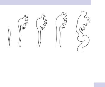
Grade III or more may require surgical intervention, and many urologists would recommend ureteric reimplantation or a STING procedure (see Fig. 7.1 for grading system for reflux).
VESICOURETERIC REFLUX (VUR) IN ADULTS 345
Grade I |
II |
III |
IV |
V |
Grade I Contrast into non-dilated ureter
Grade II Contrast into renal pelvis and calyces; no dilatation Grade III Mild dilatation of ureter; pelvis, and calyces
Grade IV Dilated ureter becomes slightly tortuous; moderate dilatation of pelvis and blunting of calyces
Grade V Severe ureteric dilatation and tortuosity; gross dilatation of pelvis and calyces
Figure 7.1 International reflux classification

346 CHAPTER 7 Miscellaneous urological diseases of kidney
Ureteropelvic junction (UPJ) obstruction in adults
Ureteropelvic junction (UPJ) obstruction is an obstruction of the proximal ureter at the junction with the renal pelvis resulting in a restriction of urine flow. It is known as pelviureteric junction obstruction (PUJO) outside of the United States.
Epidemiology
This affects more males than females (5:2 ratio). In unilateral cases, the left side is affected more often than the right; 10–15% of cases are bilateral.
Etiology
Congenital
-Intrinsic: Smooth muscle defect results in an aperistaltic segment of ureter at the UPJ.
-Extrinsic: compression from lower renal pole vessel over which the UPJ runs. It is unlikely that these vessels are the primary cause of the obstruction. It is more probable that UPJ obstruction leads to a dilated UPJ and ballooning of the renal pelvis over the lower pole
vessels, which may contribute to, but is not the primary cause of, the obstruction.
-Acquired: UPJ stricture secondary to ureteral manipulation (e.g., ureteroscopy); trauma from passage of calculi; fibroepithelial polyps; TCC of urothelium at UPJ; external compression of ureter by retroperitoneal fibrosis or malignancy
Presentation
Patients present with flank pain precipitated by a diuresis (high fluid intake; especially precipitated by consumption of alcohol), flank mass, UTI, and hematuria (after minor trauma). It may also be associated with vesicoureteric reflux (VUR).
Investigation
Renal ultrasound shows renal pelvis dilatation in the absence of a dilated ureter. IVP demonstrates delay of excretion of contrast and a dilated pelvicalyceal system. Arrange for a CT, to exclude a small, radiolucent stone, urothelial TCC, or retroperitoneal pathology that may be the cause of the obstruction at the UPJ.
MAG3 renography with administration of Lasix to establish a maximum diuresis is the definitive diagnostic test for UPJ obstruction. Radioisotope accumulates in the renal pelvis, and following IV Lasix it continues to accumulate (a rising curve).
Many urologists perform retrograde pyelography to establish the exact site of the obstruction, but they do this at the time of UPJ repair to avoid introducing infection into an obstructed renal pelvis.