Материал: Oxford American Handbook of Urology ( PDFDrive )

URETEROPELVIC JUNCTION (UPJ) OBSTRUCTION IN ADULTS 347
Treatment
Surgery
Surgery is indicated for recurrent episodes of bothersome pain, renal impairment, when a stone has developed in the obstructed kidney, and when infection (pyonephrosis) has supervened.
In the absence of symptoms, consider watchful waiting with serial MAG3 renograms. If renal function remains stable and the patient remains free of symptoms, there is no need to operate.
Endoscopic treatment of a UPJ obstruction is called an endopyelotomy (or pyelolysis). Various techniques have been described, but the essential principle is the same—full-thickness incision through the obstructing proximal ureter, from within the lumen of the ureter down into the peripelvic and periureteral fat, using a sharp knife or holmium:YAG laser. The incision is stented for 4 weeks to allow re-epithelialization of the UPJ. The procedure is relatively minimally invasive and is generally not used for UPJ obstruction >2 cm in length.
The incision may be made percutaneously or by a retrograde approach via a rigid or flexible ureteroscope, or by using a specially designed endopyelotomy balloon (the Acucise® technique). An angioplasty-type balloon over which a cautery wire runs is inflated across the UPJ. Passage of an electrical current heats the wire and this cuts through the obstructing ring of tissue at the UPJ.
The presence of a combination of UPJ obstruction and a renal stone that is suitable for PCNL is an indication for combined PCNL and percutaneous endopyelotomy.
Success rates in terms of relieving obstruction are as follows: percutaneous endopyelotomy, 60–100% (mean 70%); cautery wire balloon endopyelotomy, 70%; ureteroscopic endopyelotomy, 80%.
Pyeloplasty
-Open pyeloplasty has success rates of 95% and may also be used after endopyelotomy failure or as a first-line technique.
-Laparoscopic or robotic-assisted pyeloplasty has the advantage of accelerated patient recovery, with success rates of 95%.
•Common techniques include dismembered pyeloplasty (also known as the Anderson–Hynes pyeloplasty: the narrowed area of UPJ is excised, the proximal ureter is spatulated and anastomosed to the renal pelvis), flap pyeloplasty (Culp), and Y-V-plasty (Foley).

348 CHAPTER 7 Miscellaneous urological diseases of kidney
Anomalies of renal ascent and fusion: horseshoe kidney, pelvic kidney, malrotation
Abnormalities of renal ascent and fusion occur in weeks 6–9 of gestation, when the embryonic kidney is ascending to its definitive lumbar position (ascending as a result of rapid caudal growth of the embryo).
Horseshoe kidney
This is the most common example of renal fusion. Prevalence is 1 in 400, with a male-to-female ratio 2:1.
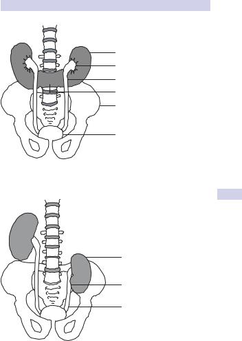
The kidneys lie vertically (instead of obliquely) and are joined at their lower poles (in 95%) by midline parenchymal tissue (the isthmus). The inferior mesenteric artery obstructs ascent of the isthmus. Consequently, the horseshoe kidney lies lower in the abdomen (at L3 or L4 vertebral level).
Normal rotation of the kidney is also prevented, thus the renal pelvis lies anteriorly, with the ureters also passing anteriorly over the kidneys and isthmus (but entering the bladder normally). Blood supply is variable, usually from one or more renal arteries or their branches, or from branches off the aorta or inferior mesenteric artery (see Fig. 7.2).
A proportion of individuals with horseshoe kidneys have associated congenital abnormalities (Turner syndrome, trisomy 18, genitourinary anomalies, ureteric duplication), vesicoureteric reflux, UPJ obstruction, and renal tumors (including Wilm tumors).
Most patients with horseshoe kidneys remain asymptomatic; however, infection and calculi may develop and cause symptoms. The diagnosis is usually suggested on renal ultrasound and confirmed by IVP (calyces of the lower renal pole are seen to point medially, and lie medially in relation to the ureters) or CT. Renal function is usually normal.
Pelvic kidney
This represents a form of renal ectopia. Prevalence is 1 in 2000–3000, with both sexes affected equally.
The left kidney is affected more often than the right, and bilateral cases are seen in <10%. The affected kidney is smaller, with the renal pelvis positioned anteriorly (instead of medially), and the ureter is short but enters the bladder normally.
Pelvic kidneys lie opposite the sacrum and below the aortic bifurcation and are supplied by adjacent (aberrant) vessels (see Fig. 7.3). There is an increased risk of congenital anomalies, including contralateral renal agenesis and genital malformations.
Most cases are asymptomatic. Diagnosis is made on renal ultrasound scan, IVP, or renography. Complications include obstruction, hydronephrosis, and infection.
ANOMALIES OF RENAL ASCENT AND FUSION 349
Horseshoe kidney (vertical orientation)
Anterior renal pelvis
Ureter (running anterior to kidney)
Isthmus
Bony pelvis
Bladder
Figure 7.2 Horseshoe kidney.
Smaller pelvic kidney
Anterior renal pelvis
Short ureter
Figure 7.3 Pelvic (ectopic) kidney.

350 CHAPTER 7 Miscellaneous urological diseases of kidney
Malrotation
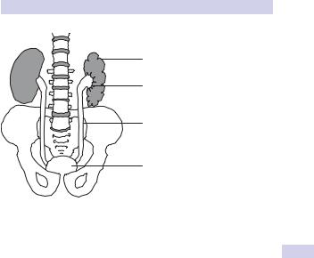
The kidney is located in a normal position, but the renal pelvis fails to rotate from an anterior to a medial orientation. Prevalence is ~1 in 1000, with a male-to-female ratio of 2:1. The renal shape may be altered (flattened, oval, triangular, or elongated) and the kidney retains its fetal lobulated outline (see Fig. 7.4).
It is associated with increased deposition of fibrous tissue around the renal hilum, which can produce symptoms due to ureteric or UPJ obstruction (causing hydronephrosis, infection, or stone formation). Most patients, however, remain asymptomatic.
The diagnosis is made on renal ultrasound scan, IVP, or retrograde pyelography.
ANOMALIES OF RENAL ASCENT AND FUSION 351
Altered renal shape (with fetal lobulations)
Anterior renal pelvis
Ureter
Bladder
Figure 7.4 Malrotation of the kidney.