Материал: Oxford American Handbook of Urology ( PDFDrive )

372 CHAPTER 8 Stone disease
Stone fragmentation techniques: extracorporeal lithotripsy (ESWL)
The technique of focusing externally generated shock waves at a target (the stone) was first used in humans in 1980. The first commercial lithotriptor, the Dornier HM3, became available in 1983.1 ESWL revolutionized kidney and ureteric stone treatment.
Three methods of shock wave generation are commercially available— electrohydraulic, electromagnetic, and piezoelectric.
Electrohydraulic
Application of a high-voltage electrical current between 2 electrodes about 1 mm apart under water causes discharge of a spark. Water around the tip of the electrode is vaporized by the high temperature, resulting in a rapidly expanding gas bubble. The rapid expansion and then rapid collapse of this bubble generates a shock wave that is focused by a metal reflector shaped as a hemi-ellipsoid.
This method was used in the original Dornier HM3 lithotriptor.
Electromagnetic
Two electrically conducting cylindrical plates are separated by a thin membrane of insulating material. Passage of an electrical current through the plates generates a strong magnetic field between them, the subsequent movement of which generates a shock wave. An acoustic lens is used to focus the shock wave.
Piezoelectric
A spherical dish is covered with about 3000 small ceramic elements, each of which expands rapidly when a high voltage is applied across them. This rapid expansion generates a shock wave. X-ray, ultrasound, or combinations of both are used to locate the stone on which the shock waves are focused.
Older machines required general or regional anesthesia because the shock waves were powerful and caused severe pain.
Newer lithotriptors generate less powerful shock waves, allowing ESWL with oral or parenteral analgesia in many cases, but they are less efficient at stone fragmentation.
Efficacy of ESWL
The likelihood of fragmentation with ESWL depends on stone size and location, anatomy of renal collecting system, degree of obesity, and stone composition. ESWL is most effective for stones <2 cm in diameter, in favorable anatomical locations. It is less effective for stones >2 cm diameter, in lower-pole stones in a calyceal diverticulum (poor drainage), and those composed of cystine or calcium oxalate monohydrate (very hard).
Stone-free rates for solitary kidney stones are 80% for stones <1 cm in diameter, 60% for those between 1 and 2 cm, and 50% for those >2 cm in diameter. Lower stone-free rates as compared with open surgery or PCNL are accepted because of the minimal morbidity of ESWL.
1 Chaussy CG, Brendel W, Schmidt E (1980). Extracorporeal induced destruction of kidney stones by shock waves. Lancet 2:1265–1268.

STONE FRAGMENTATION TECHNIQUES: ESWL 373
There have been no randomized studies comparing stone-free rates between different lithotriptors. In nonrandomized studies, rather surprisingly, when it comes to efficacy of stone fragmentation, older (the original Dornier HM3 machine) is better (but with higher requirement for analgesia and sedation or general anesthesia). Less powerful (modern) lithotriptors have lower stone-free rates and higher retreatment rates.
Side effects of ESWL
ESWL causes a certain amount of structural and functional renal damage (found more frequently the harder you look). Hematuria (microscopic, macroscopic) and edema are common, perirenal hematomas less so (0.5% detected on ultrasound with modern machines, although reported in as many as 30% with the Dornier HM3).
Effective renal plasma flow (measured by renography) has been reported to fall in ~30% of treated kidneys. There are data suggesting that ESWL may increase the likelihood of development of hypertension.
Acute renal injury may be more likely to occur in patients with preexisting hypertension, prolonged coagulation time, coexisting coronary heart disease, or diabetes and in those with solitary kidneys.
Contraindications to ESWL
Absolute contraindications are pregnancy and uncorrected blood clotting disorders (including anticoagulation).
Procedure-specific consent form: potential complications after ESWL
Common
-Bleeding on passing urine for short period after procedure
-Pain in the kidney as small fragments of stone pass after fragmentation
-UTI from bacteria released from the stone, needing antibiotic treatment.
Occasional
-Stone will not break as too hard, requiring an alternative treatment
-Repeated ESWL treatments may be required
-Recurrence of stones
Rare
-Kidney damage (bruising) or infection, needing further treatment
-Stone fragments occasionally get stuck in the tube (ureter) between the kidney and the bladder, requiring hospital admission and sometimes surgery to remove the stone fragment
-Severe infection requiring intravenous antibiotics and sometimes drainage of the kidney by a small drain placed through the back into the kidney.
Alternative therapy
Endoscopic surgery, open surgery, or observation can be used to allow spontaneous passage.

374 CHAPTER 8 Stone disease
Intracorporeal techniques of stone fragmentation (fragmentation within the body)
Electrohydraulic lithotripsy (EHL)
EHL was the first technique developed for intracorporeal lithotripsy. A high voltage applied across a concentric electrode under water generates a spark. This vaporizes water, and the subsequent expansion and collapse of the gas bubble generates a shock wave.
EHL is an effective form of stone fragmentation. The shock wave is not focused, so the EHL probe must be applied within 1 mm of the stone to optimize stone fragmentation.
EHL has a narrower safety margin than that of pneumatic, ultrasonic, or laser lithotripsy and should be kept as far away as possible from the wall of the ureter, renal pelvis, or bladder to limit damage to these structures, and at least 2 mm away from the cystoscope, ureteroscope, or nephroscope to prevent lens fracture.
Principal uses are for bladder stones (wider safety margin than in the narrower ureter).
Pneumatic (ballistic) lithotripsy
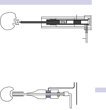
A metal projectile contained within the handpiece is propelled backward and forward at great speed by bursts of compressed air (see Fig. 8.3). It strikes a long, thin, metal probe at one end of the handpiece at 12Hz (12 strikes/second) transmitting shock waves to the probe, which, when in contact with a rigid structure such as a stone, fragments the stone.
This technique is used for stone fragmentation in the ureter (using a thin probe to allow insertion down a ureteroscope) or kidney (a thicker probe may be used, with an inbuilt suction device—Lithovac—to remove stone fragments).
Pneumatic lithotripsy is very safe, since the excursion of the end of probe is about a millimeter, and it bounces off the pliable wall of the ureter. Ureteric perforation is therefore rare.
The device is low cost and requires low maintenance. However, its ballistic effect has a tendency to cause stone migration into the proximal ureter or renal pelvis, where the stone may be inaccessible to further treatment. The metal probe cannot bend around corners, so it cannot be used for ureteroscopic treatment of stones within the kidney or with a flexible ureteroscope.
Its principal use is for ureteric stones.
Ultrasonic lithotripsy
An electrical current applied across a piezoceramic plate located in the ultrasound transducer generates ultrasound waves of a specific frequency (23,000–25,000 Hz). The ultrasound energy is transmitted to a hollow metal probe, which in turn is applied to the stone (see Fig. 8.4).
INTRACORPOREAL TECHNIQUES OF STONE FRAGMENTATION 375
Metal projectile
Probe (1–3.5mm)
Jackhammer effect
Connection to generator
Figure 8.3 The Lithoclast: a pneumatic lithotripsy device. This figure was published in Walsh PC, et al. Campbell’s Urology, 8th edition, pp. 3395–979. Copyright Elsevier 2002.
Ultrasound generator
Piezoceramic elements
Longitudinal vibration
Ultrasound |
|
probe |
Acoustical end parts |
Suction |
Foot |
|
piece |
Figure 8.4 The Calcuson: an ultrasonic lithotripsy device. This figure was published in Walsh PC, et al. Campbell’s Urology, 8th edition, pp. 3395–979. Copyright Elsevier 2002.
The stone resonates at high frequency and this causes it to break into small fragments (the opera singer breaking a glass) that are then sucked out through the center of the hollow probe. Soft tissues do not resonate when the probe is applied to them and thus are not damaged.
This technique can only be used down straight, rigid instruments. Principal uses include fragmentation of renal calculi during PCNL.

376 CHAPTER 8 Stone disease
Laser lithotripsy
The holmium:YAG laser is principally a photothermal mechanism of action, causing stone vaporization. It has minimal shock-wave generation and therefore less risk of causing stone migration. The laser energy is delivered down fibers that vary in diameter from 200 to 360 microns. The 200-micron fiber is very flexible and can be used to gain access to stones even within the lower pole of the kidney (see Figs. 8.5 and 8.6).
The zone of thermal injury is limited to 0.5–1 mm from the laser tip. No stone can withstand the heat generated by the Ho:YAG laser. Laser lithotripsy takes time; however, since the thin laser fiber must be “painted” over the surface of the stone to vaporize it.
Principal uses are for ureteric stones and small intrarenal stones.