Материал: Oxford American Handbook of Urology ( PDFDrive )
INTRACORPOREAL TECHNIQUES OF STONE FRAGMENTATION 377
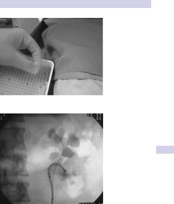
Figure 8.5 A laser fiber.
Figure 8.6 Access to the lower pole of the kidney with a flexible ureteroscope.

378 CHAPTER 8 Stone disease
Kidney stone treatment: flexible ureteroscopy and laser treatment
The development of small-caliber ureteroscopes with active deflecting mechanisms and instrument channels, in combination with the development of laser technology, small-diameter laser fibers, and stone baskets and graspers, has opened the way for intracorporeal, endoscopic treatment of kidney stones.
Access to virtually the entire collecting system is possible with modern instruments. The holmium:YAG laser has a minimal effect on tissues at distances of 2–3 mm from the laser tip and so collateral tissue damage is minimal with this laser type.
Flexible ureteroscopy and laser fragmentation offers a more effective treatment option than ESWL, with a lower morbidity than PCNL, but usually requires a general anesthetic (some patients will tolerate it with sedation alone). It can also allow access to areas of the kidney where ESWL is less efficient or where PCNL cannot reach. It is most suited to stones <2 cm in diameter.
Indications for flexible ureteroscopic kidney stone treatment
-ESWL failure
-Lower pole stone (reduces likelihood of stone passage postESWL— fragments have to pass uphill)
-Cystine stones
-Obesity such that PCNL access is technically difficult or impossible (nephroscopes may not be long enough to reach stone)
-Obesity such that ESWL is technically difficult or impossible. BMI >28 is associated with lower ESWL success rates. Treatment distance may exceed focal length of lithotriptor.
-Musculoskeletal deformities such that stone access by PCNL or ESWL is difficult or impossible (e.g., kyphoscoliosis)
-Stone in a calyceal diverticulum (accessing stones in small diverticulae in upper and anterior calyces is difficult and carries significant risks)
-Stenosis of a calyceal infundibulum or tight angle between renal pelvis and infundibulum. The flexible ureteroscope can negotiate acute angles and the laser can be used to divide obstructions.
-Bleeding diathesis where reversal of this diathesis is potentially dangerous or difficult
-Horseshoe or pelvic kidney. ESWL fragmentation rates are only 50% in such cases1 due to difficulties of shock-wave transmission through overlying organs (bowel). PCNL for such kidneys is difficult because of bowel proximity and variable blood supply (blood supply derived from multiple sources).
-Patient preference
1 Kupeli B, Isen K, Biri H, et al. (1999). Extracorporeal shockwave lithotripsy in anomalous kidneys.
J Endourol 13:39–52.

KIDNEY STONE TREATMENT 379
Disadvantages
Efficacy diminishes as stone burden increases—it simply takes a long time to paint the surface of the stone with laser energy, causing fragmentation.
A dust cloud is produced as the stone fragments, and this temporarily obscures the view until it has been washed away by irrigation. Stone fragmentation rates for those expert in flexible ureteroscopy are ~70–80% for stones <2 cm in diameter and 50% for those >2 cm in diameter,2 and ~10% of patients will require two or more treatment sessions.
2 Dasgupta P, et al. (2004). Flexible ureterorenoscopy: prospective analysis of the Guy’s experience. Ann R Coll Surg 86:367–370.

380 CHAPTER 8 Stone disease
Kidney stone treatment: percutaneous nephrolithotomy (PCNL)
Technique
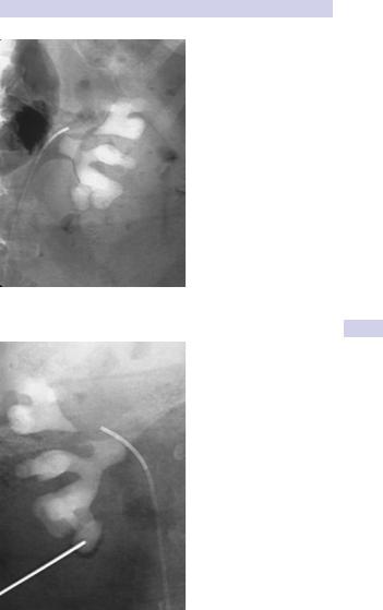
PCNL is the removal of a kidney stone via a track developed between the surface of the skin and the collecting system of the kidney. The first step requires inflation of the renal collecting system (pelvis and calyces) with fluid or air instilled via a ureteric catheter inserted cystoscopically (Fig. 8.7). This makes subsequent percutaneous puncture of a renal calyx with a nephrostomy needle easier (Fig. 8.8).
Once the nephrostomy needle is in the calyx, a guide wire is inserted into the renal pelvis to act as a guide over which the track is dilated (Fig. 8.9). An access sheath is passed down the track and into the calyx, and through this a nephroscope can be advanced into the kidney (Fig. 8.10). An ultrasonic lithotripsy probe is used to fragment the stone and remove the debris.
A posterior approach is most commonly used, below the 12th rib (to avoid the pleura and far enough away from the rib to avoid the intercostals, vessels, and nerve). The preferred approach is through a posterior calyx, rather than into the renal pelvis, because this avoids damage to posterior branches of the renal artery that are closely associated with the renal pelvis. General anesthesia is usual, though regional or even local anesthesia (with sedation) can be used.
Indications for PCNL
PCNL is generally recommended for stones >3 cm in diameter and those that have failed ESWL and/or an attempt at flexible ureteroscopy and laser treatment. It is the first-line option for staghorn calculi,1 with ESWL and/or repeat PCNL being used for residual stone fragments.
For stones 2–3 cm in diameter, options include ESWL (with a JJ stent in situ), flexible ureteroscopy and laser treatment, and PCNL. PCNL gives the best chance of complete stone clearance with a single procedure, but this is achieved at a higher risk of morbidity.
Some patients will opt for several sessions of ESWL or flexible ureteroscopy/laser treatment and the possible risk of ultimately requiring PCNL because of failure of ESWL or laser treatment, rather than proceeding with PCNL up front.
About half of stones >2 cm in diameter will be fragmented by flexible ureteroscopy and laser treatment.
Outcomes of PCNL
For small stones, the stone-free rate after PCNL is on the order of 90–95%. For staghorn stones, the stone-free rate of PCNL, combined with postoperative ESWL for residual stone fragments, is on the order of 80–85%.
1 Segura JW, Preminger GM, Assimos DG, et al. (1994). Nephrolithiasis clinical guidelines panel summary report on the management of staghorn calculi. J Urol 151:1648–1651.
KIDNEY STONE TREATMENT: PCNL 381
Figure 8.7 A ureteric catheter is inserted into the renal pelvis to dilate it with air or fluid.
Figure 8.8 A nephrostomy needle has been inserted into a calyx.