Материал: Oxford American Handbook of Urology ( PDFDrive )

TREATMENT OPTIONS FOR URETERIC STONES 397
Failed initial ESWL is associated with a low success rate for subsequent ESWL. Therefore, if ESWL has no effect after one or two treatments, change tactics.2
Open ureterolithotomy and laparoscopic ureterolithotomy are used when ESWL or ureteroscopy have been tried and failed or were not feasible.
2 Pace KT. et al. (2000). Low success rate of repeat shock wave lithotripsy for uretal stones after failed initial treatment. J Urol 164:1905–1907.

398 CHAPTER 8 Stone disease
Prevention of calcium oxalate stone formation
A series of landmark papers from Harvard Medical School1 and other groups help us to give rational advice on reducing the risk of future stone formation in those who have formed one or more stones. The Harvard studies stratified risk of stone formation on the basis of intake of calcium and other nutrients (Nurses Health Study, n = 81,000 women; equivalent male study, n = 45,000).
Low fluid intake
Low fluid intake may be the single most important risk factor for recurrent stone formation. High fluid intake is protective,1 by reducing urinary saturation of calcium, oxalate, and urate.
Time to recurrent stone formation is prolonged from 2 to 3 years in previous stone formers randomized to high fluid vs. low fluid intake (averaging about 2.5 vs. 1 L/day) and over 5 years, risk of recurrent stones was 27% in low-volume controls compared with 12% in high-volume patients.2
Dietary calcium
Conventional teaching was that high calcium intake increases the risk of calcium oxalate stone disease. The Harvard Medical School studies have shown that low calcium intake is, paradoxically, associated with an increased risk of forming kidney stones, in both men and women (relative risk of stone formation for the highest quintile of dietary calcium intake vs. the lowest quintile = 0.65; 95% confidence intervals 0.5 to 0.83—i.e., high calcium intake was associated with a low risk of stone formation).
Calcium supplements
In the Harvard studies,3 the relative risk of stone formation in women on supplemental calcium compared with those not on calcium was 1.2 (95% confidence intervals 1.02–1.4). In 67% of women on supplements, the calcium was either not consumed with a meal or was consumed with a meal with low oxalate content. It is possible that consuming calcium supplements with a meal or with oxalate-containing foods could reduce this small risk of inducing kidney stones.
Other dietary risk factors related to stone formation
Increased risk of stone formation (relative risk of stone formation shown in brackets for highest to lowest quintiles of intake of particular dietary factor):
-Sucrose [1.5]
-Sodium [1.3]: high sodium intake (leading to natriuresis) causes hypercalciuria
-Potassium [0.65]
1 Curhan GC, et al. (1993). A prospective study of dietary calcium and other nutrients and the risk of symptomatic kidney stones. N Engl J Med 328:833–38.
2 Borghi L, et al. (1996). Urinary volume, water and recurrences in idiopathic calcium nephrolithiasis: A 5-year randomized prospective study. J Urol 155:839–843.
3 Curhan G, et al. (1997). Comparison of dietary calcium with supplemental calcium and other nutrients as factors affecting the risk for kidney stones in women. Ann Intern Med 126:497–504.

PREVENTION OF CALCIUM OXALATE STONE FORMATION 399
Animal proteins
High intake of animal proteins causes increased urinary excretion of calcium, reduced pH, high urinary uric acid, and reduced urinary citrate, all of which predispose to stone formation.4
Alcohol
Curhan’s studies from Harvard5 suggest small quantities of wine decrease risk of stones.
Vegetarian diet
Vegetable proteins contain less of the amino acids phenylalanine, tyrosine, and tryptophan that increase the endogenous production of oxalate. A vegetarian diet may protect against the risk of stone formation.6,7
Dietary oxalate
A small increase in urinary oxalate concentration increases calcium oxalate supersaturation much more than an increase in urinary calcium concentration. Mild hyperoxaluria is one of the main factors leading to calcium stone formation.8
4 Kok DJ (1990). The effects of dietary excesses in animal protein and in sodium on the composition and crystallization kinetics of calcium oxalate monohydrate in urines of healthy men. J Clin Endocrinol Metab 71:861–867.
5 Curhan G, et al. (1998). Beverage use and risk for kidney stones in women. Ann Intern Med 128:534–540.
6 Robertson WG, et al. (1982). Prevalence of urinary stone disease in vegetarians. Eur Urol 8:334–339. 7 Borghi, L (2002). Comparison of two diets for prevention of recurrent stones in idiopathic hypercalciuria. N Engl J Med 346:77–84.
8 Robertson WG, Peacock M, Ouimet D, et al. (1981). The main risk for calcium oxalate stone disease in man: hypercalciuria or mild hyperoxaluria? In Smith LH, Robertson WG, Finlayson B (Eds.), Urolithiasis: Clinical and Basic Research. New York: Plenum Press, pp. 3–12.

400 CHAPTER 8 Stone disease
Bladder stones
Composition
Bladder stones consist of struvite (i.e., they are infection stones) or uric acid (in noninfected urine).
Adults
Bladder calculi are predominantly a disease of men aged >50 and with bladder outlet obstruction due to benign prostatic enlargement (BPE). They also occur in the chronically catheterized patient (e.g., spinal cord injury patients), in whom the chance of developing a bladder stone is 25% over 5 years (similar risk whether urethral or suprapubic location of the stone).1
Children
Bladder stones are still common in Thailand, Indonesia, North Africa, the Middle East, and Burma. In these endemic areas they are usually composed of a combination of ammonium urate and calcium oxalate. A low-phos- phate diet in these areas (a diet of breast milk and polished rice or millet) results in high peaks of ammonia excretion in the urine.
Symptoms
Bladder stones may be symptomless (incidental finding on KUB X-ray or bladder ultrasound or on cystoscopy) and is the common presentation in spinal patients who have limited or no bladder sensation.
In the neurologically intact patient, suprapubic or perineal pain, hematuria, urgency and/or urge incontinence, recurrent UTI, and LUTS (hesitancy, poor flow) may occur.
Diagnosis
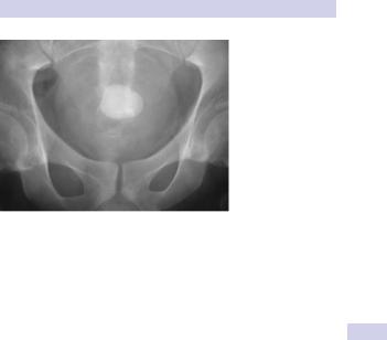
If you suspect a bladder stone, it will be visible on KUB X-ray or renal ultrasound (Fig. 8.15).
Treatment
Most stones are small enough to be removed cystoscopically (endoscopic cystolitholapaxy), using stone-fragmenting forceps for stones that can be engaged by the jaws of the forceps and EHL or pneumatic lithotripsy for those that cannot. Large stones (see Fig. 8.15) can be removed by open surgery (open cystolitholapaxy).
1 Ord J (2003). Bladder management and risk of bladder stone formation in spinal cord injured patients. J Urol. 170:1734–1737.
BLADDER STONES 401
Figure 8.15 A bladder stone.