Материал: Oxford American Handbook of Urology ( PDFDrive )

422 CHAPTER 10 Trauma to the urinary tract
Renal trauma: clinical and radiological assessment
Renal injuries may not initially be obvious, hidden as they are by other structures. To confirm or exclude a renal injury, imaging studies are required. In children, there is proportionately less perirenal fat to cushion the kidneys against injury, thus renal injuries occur with lesser degrees of trauma. Staging of renal injuries is given in Box 10.1.
History includes mechanism of the trauma (blunt, penetrating).
Examination
Pulse rate, systolic blood pressure, respiratory rate, location of entry and exit wounds, flank bruising, and rib fractures need to be assessed. The lowest recorded systolic blood pressure is used to determine the presence of shock and thus the need for renal imaging.
Remember, in young adults and children, hypotension is a late manifestation of hypovolemia; blood pressure is maintained until there has been substantial blood loss, thus making shock a less reliable indicator.
Indications for renal imaging
•Gross hematuria
•Microscopic (>5 RBCs per high-powered field [hpf]) or dipstick hematuria in a hypotensive patient (systolic blood pressure of <90 mmHg recorded at any time since the injury1)
•History of rapid deceleration with evidence of multisystem trauma (e..g., fall from a height, high-speed motor vehicle accident).
•Penetrating chest and abdominal wounds (knives, bullets) with any degree of hematuria or suspicion of renal injury based on wound location
•Any child with urinalysis showing t50 RBC/hpf after blunt trauma
Adult and pediatric patients with isolated microhematuria after blunt trauma need not have their kidneys imaged immediately as long as there is no history of rapid deceleration and no evidence of multisystem trauma or shock, since the chances of a significant renal injury are <0.2% in this setting.
Degree of hematuria
While deep renal lacerations may often be associated with obvious gross hematuria, in some cases of severe renal injury (renal vascular injury, UPJ avulsion) hematuria may be absent. Thus the relationship between the presence, absence, and degree of hematuria and the severity of renal injury is neither predictable nor reliable.
With blunt renal trauma in adults there is a chance of significant renal injury vs. degree of hematuria and systolic blood pressure (Table 10.1).

RENAL TRAUMA: CLINICAL AND RADIOLOGICAL ASSESSMENT 423
Box 10.1 Staging of the renal injury
Using CT, renal injuries are staged according to the American Association for the Surgery of Trauma (AAST) Organ Injury Severity Scale. Higher injury severity scales are associated with poorer outcomes.
Grade I Contusion or subcapsular hematoma with no parenchymal laceration
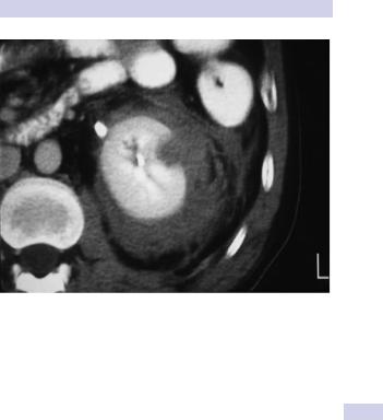
Grade II Parenchymal laceration of cortex <1 cm deep, no extravasation of urine (i.e., collecting system intact) (Fig. 10.1)
Grade III Parenchymal laceration of cortex >1 cm deep, no extravasation of urine (i.e., collecting system intact)
Grade IV Parenchymal laceration involving cortex, medulla, and collecting system OR segmental renal artery or renal vein injury with contained hemorrhage
Grade V Completely shattered kidney OR avulsion of renal hilum
Table 10.1 Renal injury as indicated by hematuria and SBP
Degree of hematuria; systolic BP (mmHg) |
Significant renal injury |
Microhematuria;* SBP >90 |
0.2% |
Gross hematuria; SBP >90 |
10% |
Gross or microhematuria; SBP <90 |
10% |
|
|
* Dipstick or microscopic hematuria

424 CHAPTER 10 Trauma to the urinary tract
The hemodynamically unstable patient
While CT is always the preferred imaging method for stable patients with suspected renal injuries, hemodynamic instability may preclude its use. Such patients may need to be taken to the operating room immediately to control bleeding. In this situation, an intraoperative on-table IVP (see Table 10.1) is indicated if
•A retroperitoneal hematoma is found and/or
•A renal injury is found that is likely to require nephrectomy.
RENAL TRAUMA: CLINICAL AND RADIOLOGICAL ASSESSMENT 425
Figure 10.1 Renal CT with IV contrast in blunt trauma patent shows a superficial (grade 2) laceration amenable to nonoperative management.

426 CHAPTER 10 Trauma to the urinary tract
Renal trauma: treatment
Conservative (nonoperative) management
Most blunt (95%) and many penetrating renal injuries (50% of stab injuries and 25% of gunshot wounds) can be managed nonoperatively.
Dipstick or microscopic hematuria: If systolic BP since injury has always been >90 mmHg and there is no history of deceleration, imaging and admission is not required. Outpatient follow-up of microhematuria should be considered.
Gross hematuria: In a hemodynamically stable patient whose injury has been staged with CT, admit for bed rest, antibiotics, serial labs, and observation until the hematuria resolves (cross-match in case blood pressure drops). High-grade injuries can be managed nonoperatively if they are cardiovascularly stable. However, grade IV and, especially, grade V injuries may require prompt nephrectomy to control bleeding (grade V injuries function poorly if repaired).
Surgical exploration (see Box 10.2)
This is indicated (whether blunt or penetrating injury) if
•Expanding, large, or pulsatile perirenal hematoma is present (suggests a renal pedicle avulsion; hematuria is absent in 20%).
•The patient develops shock that does not respond to resuscitation with fluids and/or blood transfusion.
•The hemoglobin decreases (there are no strict definitions of what represents a significant fall in hemoglobin).
•There is urinary extravasation and associated bowel or pancreatic injury.
Urinary extravasation
This is not an absolute indication for exploration. Almost 80–90% of these injuries will heal spontaneously. The threshold for operative repair is lower with associated bowel or pancreatic injury—bowel contents mixing with urine is a recipe for sepsis. In these situations, the renal repair should be well drained and omentum interposed between the kidney and bowel or pancreas.
If there is substantial contrast extravasation, consider placing a JJ stent and a Foley catheter.
Repeat CT imaging if the patient develops a prolonged ileus or a fever, since these signs may indicate development of a urinoma, which can be drained percutaneously. Renal exploration is needed for a persistent leak.
Devitalized segments
Exploration is usually not required for patients with devitalized segments of kidney (Fig. 10.2). If urinary extravasation is also present, these patients may be at higher risk for septic complications.