Материал: Oxford American Handbook of Urology ( PDFDrive )

RENAL TRAUMA: TREATMENT 427
Box 10.2 Technique of renal exploration
Midline incision allows the following:
•Exposure of renal pedicle, for early control of renal artery and vein
•Inspection for injury to other organs
Lift the small bowel upward to allow access to the retroperitoneum. Incise the peritoneum over the aorta, above the inferior mesenteric artery. A large perirenal hematoma may obscure the correct site for this incision. If this is the case, look for the inferior mesenteric vein and make your incision medial to this. Once on the aorta, trace it upward toward the crossing of the left renal vein. Here, both renal arteries may be accessed and vessel loops passed around these vessels.
Expose kidney by reflecting the colon up off of the retroperitoneum. Bleeding may be reduced by applying pressure to vessels via a Rummel tourniquet. Control bleeding vessels within the kidney with 4-0 absorbable sutures. Close any defects in the collecting system similarly.
If your sutures cut out, place perirenal fat or a strip of gelatin or collagen over the site of bleeding, place your sutures through the renal capsule on either side of this, and tie them over the bolster. This will stop them from cutting through the friable renal parenchyma.
Finding a nonexpanding, nonpulsatile retroperitoneal hematoma at laparotomy
An expanding or pulsatile retroperitoneal hematoma found at laparotomy in an unstable patient often indicates renal pedicle avulsion or laceration. Nephrectomy may be required to stop life-threatening hemorrhage.
Controversy surrounds management of the nonexpanding, nonpulsatile retroperitoneal hematoma found at laparotomy. If the patient is stable, most can be left alone or treated with percutaneous angiographic embolization if needed postoperatively. In inexperienced hands, renal exploration may release retroperitoneal tamponade, thus increasing risk of bleeding that can be controlled only by nephrectomy.
Preoperative or |
Action |
intraoperative |
|
imaging |
|
|
|
Normal |
Leave the hematoma alone. |
Abnormal |
Explore and repair kidney if major injury is suspected |
|
(especially for penetrating injury). Leave hematoma |
|
alone unless pulsatile and/or patient is unstable |
|
(especially blunt injuries). |
None |
Consider 1-shot IVP on table. Explore and repair renal |
|
injury if hematoma is pulsatile and patient is unstable. |
428 CHAPTER 10 Trauma to the urinary tract
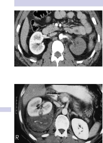
Figure 10.2 Left renal artery thrombosis after blunt trauma resulting in devitalized parenchyma successfully treated nonoperatively.
Figure 10.3 Contrast CT after abdominal stab wound shows deep central renal laceration and large perirenal hematoma with intravascular contrast extravasation. This patient remained unstable after 3 units of blood were transfused and thus underwent nephrectomy. Notice the normal contralateral kidney on this scan.

RENAL TRAUMA: TREATMENT 429
Nephrectomy
For severe renal injuries producing life-threatening bleeding, prompt nephrectomy is warranted. These are usually unstable patients who persist in shock despite multiple transfusions and have deep renal lacerations near the hilum (Fig. 10.3).
Hypertension and renal injury
Excess renin excretion occurs following renal ischemia from renal artery injury or thrombosis or renal compression by hematoma or fibrosis. This can lead to hypertension months or years after renal injury. The exact incidence of post-traumatic hypertension is uncertain but felt to be rare.
Iatrogenic renal injury: renal hemorrhage after percutaneous nephrolithotomy
Significant renal injuries can occur during percutaneous nephrolithotomy (PCNL) for kidney stones. This is the surgical equivalent of a stab wound and serious hemorrhage results in ~1% of cases.
Bleeding during or after PCNL can occur from vessels in the nephrostomy track itself, from an arteriovenous fistula, or from a pseudoaneurysm that has ruptured. Track bleeding will usually tamponade around a largebore nephrostomy tube.
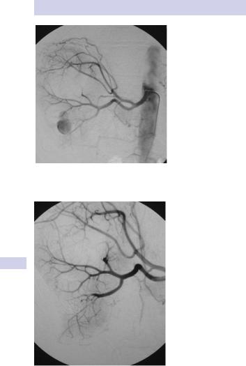
Traditionally, persistent bleeding through the nephrostomy tube is managed by clamping the nephrostomy tube and waiting for the clot to tamponade the bleeding. While this may control bleeding in some cases, in others a rising or persistently elevated pulse rate (with later hypotension) indicates the possibility of persistent bleeding and is an indication for renal arteriography and embolization of the arteriovenous fistula or pseudoaneurysm (Figs. 10.4 and 10.5). Failure to stop the bleeding by this technique is an indication for renal exploration.
Arteriovenous fistulae can sometimes occur following open renal surgery for stones or tumors, and arteriography with embolization can also be used to stop the bleeding in these cases. However, the bleeding usually occurs over a longer time course (days or even weeks), rather than as acute hemorrhage causing shock.
1 Martin X (2000). Severe bleeding after nephrolithotomy: results of hyperselective embolization. Eur Urol 37:136–139.
430 CHAPTER 10 Trauma to the urinary tract
Figure 10.4 Renal arteriography after PCNL where severe bleeding was encountered. An arteriovenous fistula was found and embolized.
Figure 10.5 Post-embolization of arteriovenous fistula. Note the embolization coils in the lower pole.
This page intentionally left blank