Материал: part17

Внимание! Если размещение файла нарушает Ваши авторские права, то обязательно сообщите нам


DICOM PS3.17 2020a - Explanatory Information​

Page 531​


...


= 4


Rows

(0028,0010)


= 4


Columns

(0028,0011)


= DIGITAL_DETECTOR


X-Ray Receptor Type

(0018,9420)


Detector Binning

(0018,701A)

= 2.0\2.0


Detector Element Spacing

(0018,7022)

= 0.2\0.2


Position of Isocenter Projection

(0018,9430)

= 5\7


...


Per-Frame Functional Groups Sequence

(5200,9230)


...


Item i


>Field of View Sequence

(0018,9432)


Item 1


>>Field of View Shape

(0018,1147)

= RECTANGLE


>>Field of View Dimensions in Float

(0018,9461)

= 1.6\1.6


>>Field of View Origin

(0018,7030)

= 2.0\2.0


>>Field of View Rotation

(0018,7032)

= 0


>>Field of View Horizontal Flip

(0018,7034)

= NO


>Frame Pixel Data Properties Sequence

(0028,9443)


Item 1


>>Imager Pixel Spacing

(0018,1164)

= 0.4\0.4


...


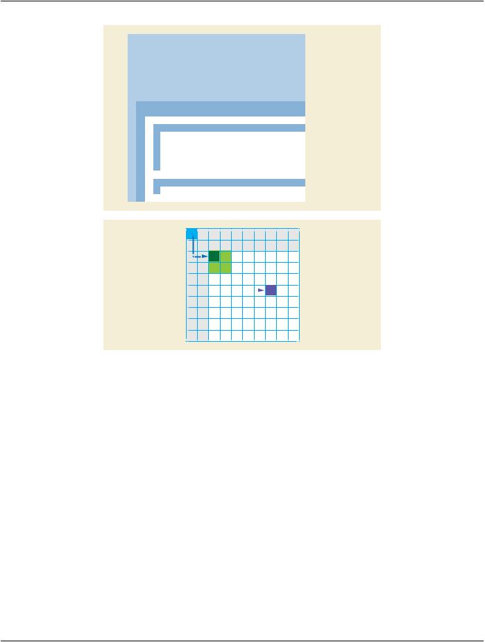
Figure FFF.2.1-27. Attributes of the Second Example of Field of View on Digital Detector​

In the third example, in addition to the binning factor of 2 between the detector matrix and the digital image, there is a resizing of 0.5​ (downsizing) between the digital image (binned) and the stored image.​

The encoded values of the key Attributes of this example are shown in Figure FFF.2.1-28.​

- Standard -​

Page 532​


DICOM PS3.17 2020a - Explanatory Information​


...


= 2


Rows

(0028,0010)


= 2


Columns

(0028,0011)


= DIGITAL_DETECTOR


X-Ray Receptor Type

(0018,9420)


Detector Binning


(0018,701A)

= 2.0\2.0


Detector Element Spacing

(0018,7022)

= 0.2\0.2


Position of Isocenter Projection

(0018,9430)

= 5\7


...


Per-Frame Functional Groups Sequence

(5200,9230)


...


Item i


>Field of View Sequence

(0018,9432)


Item 1


>>Field of View Shape

(0018,1147)

= RECTANGLE


>>Field of View Dimensions in Float

(0018,9461)

= 1.6\1.6


>>Field of View Origin

(0018,7030)

= 2.0\2.0


>>Field of View Rotation

(0018,7032)

= 0


>>Field of View Horizontal Flip

(0018,7034)

= NO


>Frame Pixel Data Properties Sequence

(0028,9443)


Item 1


>>Imager Pixel Spacing

(0018,1164)

= 0.8\0.8


...


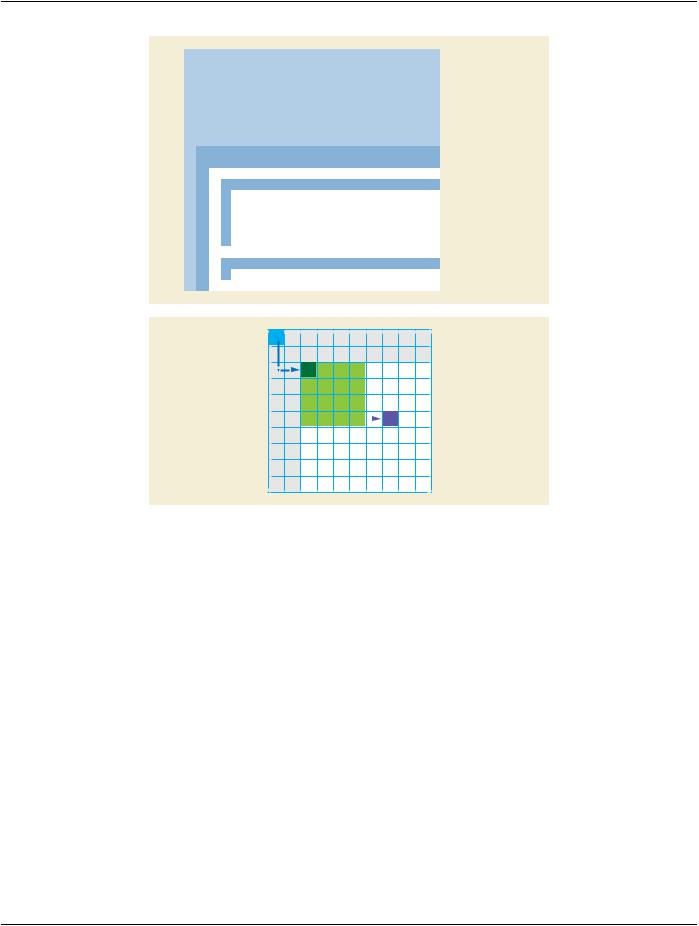
Figure FFF.2.1-28. Attributes of the Third Example of Field of View on Digital Detector​

Note that the description of the field of view Attributes (dimension, origin) is the same in these three examples. The field of view​ definition is independent from the binning and resizing processes.​

FFF.2.1.6 Acquisitions With Contrast​

ThissectionprovidesinformationontheencodingofthepresenceandtypeofcontrastbolusadministeredduringtheX-Rayacquisition.​

FFF.2.1.6.1 User Scenario​

The user performs image acquisition with injection of contrast agent during the X-Ray acquisition. Some frames are acquired without​ contrast, some others with contrast.​

The type of contrast agent can be radio-opaque (e.g., iodine) or radio-transparent (e.g., CO2).​

The information of the type of contrast and its presence or absence in the frames can be used by post-processing applications to set​ up e.g., vessel detection or image quality algorithms automatically.​

FFF.2.1.6.2 Encoding Outline​

The Enhanced XA SOP Class encodes the characteristics of the contrast agent(s) used during the acquisition of the image, including​ the type of absorption (radio-opaque or radio-transparent).​

The Enhanced XA SOP Class also allows encoding the presence of contrast in a particular frame or set of frames, by encoding the​ Contrast/Bolus Usage per-frame.​

- Standard -​

DICOM PS3.17 2020a - Explanatory Information​

Page 533​

FFF.2.1.6.3 Encoding Details​

This section provides detailed recommendations of the key Attributes to address this particular scenario.​

Table FFF.2.1-46. Enhanced X-Ray Angiographic Image IOD Modules​

IE​

Module​

PS3.3 Reference​

Usage​

Image​

Enhanced Contrast/Bolus​

C.7.6.4b​

Specifies the characteristics of the contrast​


agent(s) administered.​

Table FFF.2.1-47. Enhanced XA Image Functional Group Macros​

Functional Group Macro​

PS3.3 Reference​

Usage​

Contrast/Bolus Usage​

C.7.6.16.2.12​

Specifiesthepresenceofcontrastintheframe(s).​

FFF.2.1.6.3.1 Enhanced Contrast/bolus Module Recommendations​

The usage of this module is recommended to specify the type and characteristics of the contrast agent administered.​

FFF.2.1.6.3.2 Contrast/bolus Usage Macro Recommendations​

The usage of this macro is recommended to specify the characteristics of the contrast per-frame.​

Table FFF.2.1-48. Contrast/Bolus Usage Macro Recommendations​

Attribute Name​

Tag​

Comment​

Contrast/Bolus Usage Sequence​

(0018,9341)​

One item per contrast agent used in this frame.​

>Contrast/Bolus Agent Number​

(0018,9337)​

Contains the internal number of the agent administered as specified​


in the Enhanced Contrast/Bolus Module.​

>Contrast/Bolus Agent Administered​

(0018,9342)​

The value "YES" indicates that the contrast may be visible on the​


frame, but not necessarily if the frame is acquired before the contrast​


reaches the imaged region.​

>Contrast/Bolus Agent Detected​

(0018,9343)​

The value "YES" is used if the contrast is visible on that particular​


frame.​

Note that it is not expected to be YES if Contrast/Bolus Agent​

Administered (0018,9342) equals NO.​

FFF.2.1.6.4 Example​

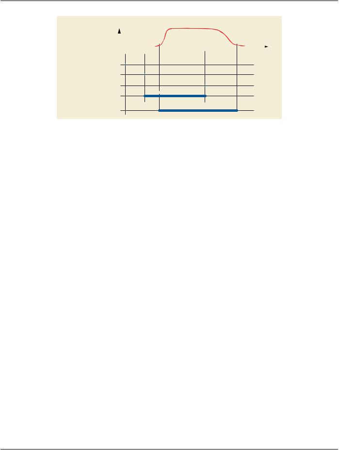
In this example, the user starts the X-Ray acquisition at 4 frames per second at 3:35pm. After one second the user starts the injection​ of 45 milliliters of contrast media Iodipamide (350 mg/ml Cholographin (Bracco) ) at a flow rate of 15 ml/sec during three seconds, in​ intra-arterial route. When the injection of contrast agent is finished, the user continues the X-Ray acquisition for two seconds until​ wash out of the contrast agent.​

There could be two ways to determine the presence of contrast agent on the frames:​

•​The injector is connected to the X-Ray acquisition system, the presence of contrast agent is determined based on the injector​ start/stop signals and a preconfigured delay to allow the contrast to reach the artery of interest, or.​

•​The X-Ray system processes the images in real time and detects the presence or absence of contrast agent on the images.​

In this example, the image acquired contains 25 frames: From frames 5 to 17, the contrast is being injected. From frames 8 to 23,​ the contrast is visible on the pixel data.​

The figure below shows the Attributes of this example in a graphical representation of the multi-frame acquisition.​

- Standard -​

Page 534​

DICOM PS3.17 2020a - Explanatory Information​

Contrast/bolus intensity visible on the pixel data


1

5


Frames


time


0

1

2

3

4

5

6


Seconds

Acquisition DateTime (0008,002A)

Contrast/Bolus Start Time (0018,1042)

Contrast/Bolus Stop Time (0018,1043)

Injection of contrast agent

Injection of contrast agent

Contrast Flow Duration (0018,1047)

Contrast agent visible on the pixels

Contrast agent visible on the pixels

Figure FFF.2.1-29. Example of contrast agent injection​

The encoded values of the key Attributes of this example are shown in Figure FFF.2.1-30.​

- Standard -​

DICOM PS3.17 2020a - Explanatory Information​

Page 535​

Contrast/Bolus Agent Sequence

(0018,0012)


Contrast agent 1


Item 1


>Code Value

(0008,0100)

= 73212002


>Coding Scheme Designator

(0008,0102)


= SCT


>Code Meaning

(0008,0104)


= Iodipamide


>Contrast/Bolus Agent Number

(0018,9337)

= 1


>Contrast/Bolus Administration Route Sequence

(0018,0014)


Item 1


>>Code Value

(0008,0100)

= 58100008


>>Coding Scheme Designator

(0008,0102)


= SCT


>>Code Meaning

(0008,0104)


= Intra-arterial route


>Contrast/Bolus Ingredient Code Sequence

(0018,9338)


Item 1


>>Code Value

(0008,0100)

= 44588005


>>Coding Scheme Designator

(0008,0102)


= SCT


>>Code Meaning

(0008,0104)


= Iodine


>Contrast/Bolus Volume

(0018,1041)

= 45


>Contrast/Bolus Ingredient Concentration

(0018,1049)

= 350


>Contrast/Bolus Ingredient Opaque

(0018,9425)


= YES


>Contrast Administration Profile Sequence

(0018,9340)


Item 1


Administered phase 1


>>Contrast/Bolus Volume

(0018,1041)

= 45


>>Contrast/Bolus Start Time

(0018,1042)

= 001536.0


>>Contrast/Bolus Stop Time

(0018,1043)

= 001539.0


>>Contrast Flow Rate

(0018,1046)

= 15


>>Contrast Flow Duration

(0018,1047)

= 3


...


Per-Frame Functional Groups Sequence

(5200,9230)


Items 1 to 4


Frames 1 to 4


>Contrast/Bolus Usage Sequence

(0018,9341)


Item 1


>>Contrast/Bolus Agent Number

(0018,9337)

= 1


>>Contrast/Bolus Agent Administered

(0018,9342)


= NO


>>Contrast/Bolus Agent Detected

(0018,9343)


= NO


Items 5 to 7


Frames 5 to 7


>Contrast/Bolus Usage Sequence

(0018,9341)


Item 1


>>Contrast/Bolus Agent Number

(0018,9337)

= 1


>>Contrast/Bolus Agent Administered

(0018,9342)


= YES


>>Contrast/Bolus Agent Detected

(0018,9343)


= NO


Items 8 to 23


Frames 8 to 23


>Contrast/Bolus Usage Sequence

(0018,9341)


Item 1


>>Contrast/Bolus Agent Number

(0018,9337)

= 1


>>Contrast/Bolus Agent Administered

(0018,9342)


= YES


>>Contrast/Bolus Agent Detected

(0018,9343)


= YES


Items 24 to 25


Frames 24 to 25


>Contrast/Bolus Usage Sequence

(0018,9341)


Item 1


>>Contrast/Bolus Agent Number

(0018,9337)

= 1


>>Contrast/Bolus Agent Administered

(0018,9342)


= YES


>>Contrast/Bolus Agent Detected

(0018,9343)


= NO


Figure FFF.2.1-30. Attributes of Contrast Agent Injection​

FFF.2.1.7 Acquisition Parameters For X-Ray Generation (kVp, mA, …)​

This section provides information on the encoding of the parameters related to the X-Ray generation.​

FFF.2.1.7.1 User Scenario​

The user performs X-Ray acquisitions during the examination. Some of them are dynamic acquisitions where the positioner and/or​ the table have moved between frames of the Multi-frame Image, the acquisition parameters such as kVp, mA and pulse width may​ change per-frame to be adapted to the different anatomy characteristics.​

Later quality assurance wants to assess the X-Ray generation techniques in order to understand possible degradation of image​ quality, or to estimate the level of irradiation at different skin areas and body parts examined.​

- Standard -​

Источник: https://studfile.net/preview/14585770/