Материал: part17

Внимание! Если размещение файла нарушает Ваши авторские права, то обязательно сообщите нам

DICOM PS3.17 2020a - Explanatory Information​

Page 681​

projections is considered as original (i.e., the Value 1 of the Attributes Image Type (0008,0008) and Frame Type (0008,9007) equals​ ORIGINAL).​

Note that the original 2D projections can be stored as DICOM instances, and the X-Ray 3D Angiographic image can be created from​ a later reconstruction on a different equipment. In this case, since the source data is the same original set of projections, the 3D data​ is still considered as original.​

TTT.2 Application Cases​

This chapter describes different scenarios and application cases where the 3D volume is reconstructed from rotational angiography.​ Each application case is structured in four sections:​

1.​User Scenario: Describes the user needs in a specific clinical context, and/or a particular system configuration and equipment​ type.​

2.​Encoding Outline: Describes the X-Ray 3D Angiographic Image SOP Class related to this scenario, and highlights key aspects.​

3.​Encoding Details: Provides detailed recommendations of the key Attributes of the Image IOD(s) to address this particular​ scenario. The tables are similar to the IOD tables of the PS3.3. Only Attributes with a specific recommendation in this particular​ scenario have been included.​

4.​Example: Presents a typical example of the scenario, with realistic sample values, and gives details of the encoding of the key​ Attributes of the Image IOD(s) to address this particular scenario. In the values of the Attributes, the text in bold face indicates​ specific Attribute values; the text in italic face gives an indication of the expected value content.​

The first application case describes the most general reconstruction scenario, and can be considered as a baseline. The further ap-​ plication cases only describe the specificities of the new scenario vs. the baseline.​

TTT.2.1Case#1:OneRotation,One2DInstance,OneReconstruction,OneX-Ray3DInstance​

This application case is related to the most general reconstruction of a 3D volume directly from all the frames of a rotational 2D pro-​ jection acquisition.​

TTT.2.1.1 User Scenario​

The image acquisition system performs a rotational acquisition around the patient and a volume is reconstructed from the acquired​ data (e.g., through "back-projection" algorithm). The reconstruction can either occur on the same system (e.g., Acquisition Modality)​ or a secondary processing system (e.g., Co-Workstation).​

The reconstructed Volume needs to be encoded and kept saved for interchange with 3D rendering application or further equipment​ involved during an interventional procedure.​

TTT.2.1.2 Encoding Outline​

This is the basic use case of X-Ray 3D Angiographic image encoding.​

The rotational acquisition can be encoded either as a multifamily XA Image with limited frame-specific Attributes or as an Enhanced​ XA Image, with frame-specific Attributes encoded that support the algorithms to reconstruct volume data.​

The volume data is encoded as an X-Ray 3D Angiographic instance. The volume data typically spans the complete region of the​ projected matrix size (in number of rows and columns).​

All the projections of the original XA instance or Enhanced XA instance are used to reconstruct the volume.​

The X-Ray 3D Angiographic instance references the original XA instance or Enhanced XA instance and uses Attributes to define the​ context on how the original 2D image frames are used to create the volume.​

- Standard -​

Page 682​

DICOM PS3.17 2020a - Explanatory Information​

XA 2D Projection SOP Instance “C”

Contrast Frames

1...

...N

Acquisition Context #1

Reconstruction #1

X-Ray 3D SOP Instance “Z”

Contrast Volume

1...

...M

Reconstruction #1

- From Acquisition Context #1

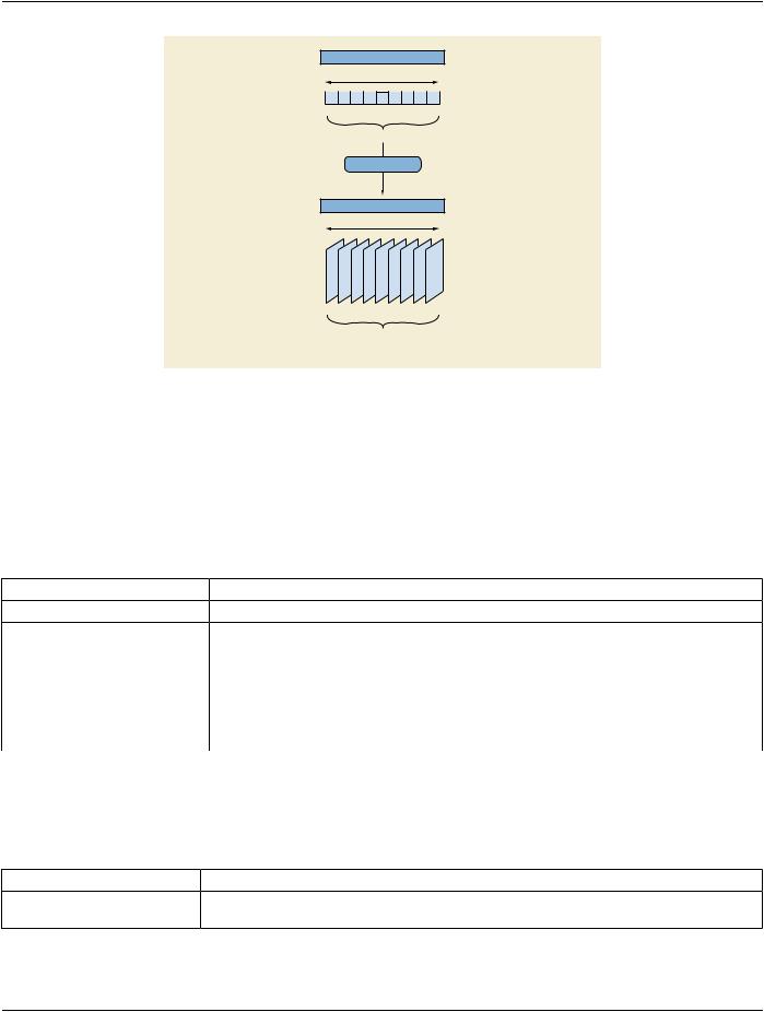
Figure TTT.2.1-1. Encoding of a 3D reconstruction from all the frames of a rotational acquisition​

TTT.2.1.3 Encoding Details​

TTT.2.1.3.1 X-Ray 3D Angiographic Image IOD​

TTT.2.1.3.1.1 General and Enhanced Series Modules Recommendations​

These modules encode the Series relationship of the created volume.​

Table TTT.2.1-1. General and Enhanced Series Modules Recommendations​

Attribute Name​

Tag​

Recommendation​

Series Instance UID​

(0020,000E)​

Use a different Series than the original projections.​

Series Description​

(0008,103E)​

Free text to describe the volume content, different from the description​


of the series of the projection images.​

Protocol Name​

(0018,1030)​

Free text to describe technical aspects of the reconstruction (focusing​


on imaging protocol rather than clinical protocol). May be relevant for​


grouping, sorting or finding of the X-Ray 3D volume.​

Referenced Performed Procedure​

(0008,1111)​

Reference to the image acquisition procedure. May also reference a​

Step Sequence​


dedicated processing procedure step (e.g., UPS).​

TTT.2.1.3.1.2 Frame of Reference Module Recommendations​

This module encodes the identifier for the spatial relationship base of this volume. If the originating 2D images do not deliver a value,​ it has to be created for the reconstructed volume.​

Table TTT.2.1-2. Frame of Reference Module Recommendations​

Attribute Name​

Tag​

Recommendation​

Frame of Reference UID​

(0020,0052)​

Volumes with identical FoR UID share the same spatial relationship. Copy​


theFoRUIDiftheoriginatingimageisencodedasanEnhancedXAImage.​

- Standard -​


DICOM PS3.17 2020a - Explanatory Information​

Page 683​

Attribute Name​

Tag​

Recommendation​


Position Reference Indicator​

(0020,1040)​

Ifthesystemiscapabletoderivesuchinformationfromtheanatomy-related​


information in the projection X-Ray image data, otherwise no​


recommendation to set a value.​


TTT.2.1.3.1.3 General and Enhanced General Equipment Modules Recommendations​

Thismoduleencodestheequipmentidentificationinformationofthesystemthatreconstructedthevolumedata.Sincethereconstruction​ is not necessarily performed by the same system that acquired the projections, the identification of the Equipment performing the re-​ construction is recommended. Furthermore the Contributing Equipment Sequence (0018,A001) of the SOP Common Module is re-​ commendedtobeusedtopreservetheidentificationofthesystemthatcreatedtheprojectionimagethatwasbaseforthereconstruction.​

TTT.2.1.3.1.4 Image Pixel Module Recommendations​

Thismoduleencodestheactualpixelsofthevolumeslices.EachsliceisencodedasoneframeoftheX-Ray3DAngiographicinstance.​ The order of the frames encoded in the pixel data is aligned with the Image Position (Patient) Attribute. The order of frames is optimal​ for simple 2D viewing if the x-,y-,z-values steadily increase or decrease.​

TTT.2.1.3.1.5 Enhanced Contrast/Bolus Module Recommendations​

This module encodes the contrast media applied. The minimum information that needs to be provided is related to the contrast agent​ and the administration route. In the reconstructed image, the contrast information comes either from the acquisition system in case​ of direct reconstruction without source DICOM instances, or from the projection images in case of reconstruction from source DICOM​ instances.​

Table TTT.2.1-3. Enhanced Contrast/Bolus Module Recommendations​

Attribute Name​

Tag​

Recommendation​

Contrast/Bolus Agent Sequence​

(0018,0012)​


>IncludeTable8.8-1“CodeSequenceMacroAttributes”inPS3.3Baseline​


See Section TTT.2.1.3.1.5.1​

CID 12 “Radiographic Contrast Agent”.​


>Contrast/Bolus Administration Route Sequence​

(0018,0014)​


>>Include Table 8.8-1 “Code Sequence Macro Attributes” in PS3.3​


See Section TTT.2.1.3.1.5.1.​

Baseline CID 11 “Route of Administration”.​


TTT.2.1.3.1.5.1 Differences between XA and Enhanced XA​

If the source instance is encoded as an Enhanced XA instance, the Enhanced Contrast/Bolus Module is specified in that IOD, then​ those values are copied from the source instance.​

If the source instance is encoded as an XA Image, only the Contrast/Bolus Module is specified in that IOD. Although acquisition​ devices are encouraged to provide details of the contrast, most of the relevant Attributes are type 3, so it is possible that if contrast​ was applied, the only indication will be the presence of Contrast/Bolus Agent (0018,0010) since that Attribute is type 2. In that case,​ if the application is unable to get more specific information from the operator, it may populate the contrast details with the generic​ (7140000, SCT, "Contrast agent") code for contrast agent and the (261665006, SCT, "Unknown") code for the administration route.​

TTT.2.1.3.1.6 Multi-frame Dimensions Module Recommendations​

This module encodes a (default) presentation order of the image frames.​

Table TTT.2.1-4. Multi-frame Dimensions Module Recommendations​

Attribute Name​

Tag​

Recommendation​

Dimension Organization Sequence​

(0020,9221)​

This will be an initial single dimension and therefore a single​


Dimension UID is sufficient.​

Dimension Organization Type​

(0020,9311)​

The value will be "3D".​

- Standard -​

Page 684​

DICOM PS3.17 2020a - Explanatory Information​

Attribute Name​

Tag​

Recommendation​

Dimension Index Sequence​

(0020,9222)​

Specifies a Dimension Index that refers to the Image Position​


(Patient) as dimension for frame order during 2D presentation of​


an X-Ray 3D volume.​

TTT.2.1.3.1.7 Patient Orientation Module Recommendations​

ThismoduleencodestheorientationofthePatientforlaterusewithsameorotherequipment.Therelatedcodedtermscanbederived​ from the Patient Position (0018,5100) according to the following table, where:​

•​PO denotes the Patient Orientation Code Sequence (0054,0410);​

•​POM denotes the Patient Orientation Modifier Code Sequence (0054,0412);​

•​PGR denotes the Patient Gantry Relationship Code Sequence (0054,0414).​

Table TTT.2.1-5. Patient Position to Orientation Conversion Recommendations​

Patient Position​

Patient Orientation Coding​

HFS​

PO: (102538003, SCT, "recumbent")​


POM: (40199007, SCT, "supine")​


PGR: (102540008, SCT, "headfirst")​

HFP​

PO: (102538003, SCT, "recumbent")​


POM: (1240000, SCT, "prone")​


PGR: (102540008, SCT, "headfirst")​

FFS​

PO: (102538003, SCT, "recumbent")​


POM: (40199007, SCT, "supine")​


PGR: (102541007, SCT, "feet-first")​

FFP​

PO: (102538003, SCT, "recumbent")​


POM: (1240000, SCT, "prone")​


PGR: (102541007, SCT, "feet-first")​

HFDR​

PO: (102538003, SCT, "recumbent")​


POM: (102535000, SCT, "right lateral decubitus")​


PGR: (102540008, SCT, "headfirst")​

HFDL​

PO: (102538003, SCT, "recumbent")​


POM: (102536004, SCT, "left lateral decubitus")​


PGR: (102540008, SCT, "headfirst")​

FFDR​

PO: (102538003, SCT, "recumbent")​


POM: (102535000, SCT, "right lateral decubitus")​


PGR: (102541007, SCT, "feet-first")​

FFDL​

PO: (102538003, SCT, "recumbent")​


POM: (102536004, SCT, "left lateral decubitus")​


PGR: (102541007, SCT, "feet-first")​

- Standard -​

DICOM PS3.17 2020a - Explanatory Information​

Page 685​

TTT.2.1.3.1.8 X-Ray 3D Image Module Recommendations​

This module encodes the specific content of the reconstructed volume.​

Table TTT.2.1-6. X-Ray 3D Image Module Recommendations​

Attribute Name​

Tag​

Recommendation​

Image Type​

(0008,0008)​

Use "ORIGINAL" value 1 (Pixel Data Characteristics) to indicate a​


reconstruction from original projections.​


Use "VOLUME" in value 3 (Image Flavor) to indicate regularly sampled.​

Icon Image Sequence​

(0088,0200)​

Includeifthereconstructionapplicationmaybeabletogeneratearendered​


representative icon image.​

TTT.2.1.3.1.9 X-Ray 3D Angiographic Image Contributing Sources Module Recommendations​

This module encodes the source SOP instances used to create the X-Ray 3D Angiographic instance.​

Table TTT.2.1-7. X-Ray 3D Angiographic Image Contributing Sources Module Recommendations​

Attribute Name​

Tag​

Recommendation​

Contributing Sources Sequence​

(0018,9506)​

One item since there is only one originating image that contributed​


to the creation of the X-Ray 3D Angiographic image.​

TTT.2.1.3.1.10 X-Ray 3D Angiographic Acquisition Module Recommendations​

This module encodes the important technical and physical parameters of the source SOP instances used to create the X-Ray 3D​ Angiographic Image instance.​

The contents of the Enhanced XA Image IOD and XA Image IOD are significantly different. Therefore the contents of the X-Ray 3D​ Acquisition Sequence will vary depending on availability of encoded data in the source instance.​

The content of the X-Ray 3D General Positioner Movement Macro provides a general overview on the Positioner data. In case a​ system does not support the Isocenter Reference System, it may still be of advantage to provide the patient-based Positioner Primary​ and Secondary Angles in the Per Projection Acquisition Sequence (0018,9538).​

The contents of the Per Projection Acquisition Sequence (0018,9538) need to be carefully aligned with the list of frame numbers in​ the Referenced Frame Numbers (0008,1160) Attribute in the Source Image Sequence (0008,2112).​

Table TTT.2.1-8. X-Ray 3D Angiographic Acquisition Module Recommendations​

Attribute Name​

Tag​

Recommendation​

X-Ray 3D Acquisition Sequence​

(0018,9507)​

One item since there is only one acquisition context that contributed​


to the reconstruction of the X-Ray 3D Angiographic image pixel data​


contents.​

TTT.2.1.3.1.11 Pixel Measures Macro Recommendations​

This module encodes the detailed size of the volume element (Pixel Spacing for row/column dimension of each slice, and Slice​ Thickness for the distance between slices). It depends on the reconstruction algorithm and is not necessarily identical to the related​ sizes in the projection images.​

For a single volume this macro is encoded "shared" as all the slices will have the same Pixel Spacing and Slice Thickness.​

TTT.2.1.3.1.12 Frame Content Macro Recommendations​

This module encodes the timing information of the frames, as well as dimension and stack index values.​

- Standard -​

Источник: https://studfile.net/preview/14585770/