Материал: part17

Внимание! Если размещение файла нарушает Ваши авторские права, то обязательно сообщите нам

DICOM PS3.17 2020a - Explanatory Information​

Page 711​

SOP Instance UID

(0008,0018)

= UID “C2”


...


Frame of Reference UID

(0020,0052)

= UID “D”


...


X-Ray Receptor Type

(0018,9420)

= DIGITAL_DETECTOR


= CARM

Positioner Type

(0018,1508)


C-Arm Positioner Tabletop Relationship

(0018,9474)

= YES


...


Per-Frame Functional Groups Sequence

(5200,9230)


...


Item i


>Isocenter Reference System Sequence

(0018,9462)


Item 1


>>Positioner Isocenter Primary Angle

(0018,9463)

=-30.0


>>Positioner Isocenter Secondary Angle

(0018,9464)

= 20.0


>>Positioner Isocenter Detector Rotation Angle

(0018,9465)

= 0.0


>>Table X Position to Isocenter

(0018,9466)

= 50.0


>>Table Y Position to Isocenter

(0018,9467)

= 30.0


>>Table Z Position to Isocenter

(0018,9468)

= 10.0


>>Table Horizontal Rotation Angle

(0018,9469)

= 0.0


>>Table Head Tilt Angle

(0018,9470)

= 0.0


>>Table Cradle Tilt Angle

(0018,9471)

= 0.0


>X-Ray Geometry Sequence

(0018,9476)


Item 1


>>Distance Source to Isocenter

(0018,9402)

= 780.0


>>Distance Source to Detector

(0018,1110)

= 1200.0

Figure TTT.2.7-8. Attributes of the Enhanced XA during the intervention​

- Standard -​

Page 712​

DICOM PS3.17 2020a - Explanatory Information​

- Standard -​

DICOM PS3.17 2020a - Explanatory Information​

Page 713​

UUUOphthalmologyUseCases(Informative)​

UUU.1 Wide Field Ophthalmic Use Cases​

Any2-dimensionalrepresentationofa3-dimensionalobjectmustundergosomekindofprojectionormappingtoformtheplanarimage.​ Within the context of imaging of the retina, the eye can be approximated as a sphere and mathematical cartography can be used to​ understandtheimpactofprojectingasphericalretinaontoaplanarimage.Whenprojectingasphericalgeometryonaplanargeometry,​ not all metric properties can be retained at the same time; some distortion will be introduced. However, if the projection is known it​ may be possible to perform calculations "in the background" that can compensate for these distortions.​

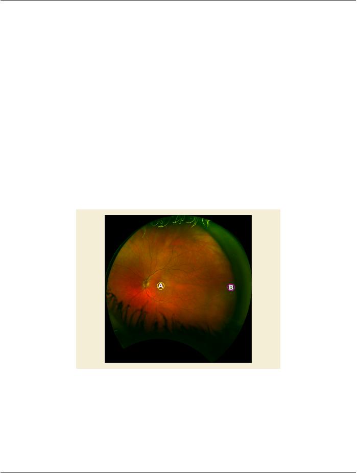
The example in Figure UUU.1-1 shows an ultra-wide field image of the human retina. The original image has been remapped to a​ stereographic projection according to an optical model of the scanning laser ophthalmoscope it was captured on. Two circles have​ been annotated with an identical pixel count. The circle focused on the fovea (A) has an area of 4.08 mm2 whereas the circle nasally​ in the periphery (B) has an area of 0.97 3mm2, both as measured with the Area Measurement using the Stereographic Projection​ method. The difference in measurement is more than 400%, which indicates how measurements on large views of the retina can be​ deceiving.​

The fact that correct measurement on the retina in physical units is difficult to do is acknowledged in the original DICOM OP SOP​ Classes in the description of the Pixel Spacing (0028,0030) tag.​

Note​

These values are specified as nominal because the physical distance may vary across the field of the images and the lens​ correction is likely to be imperfect.​

Figure UUU.1-1. Ultra-wide field image of a human retina in stereographic projection​

The following use cases are examples of how the DICOM Wide Field Ophthalmology Photography objects may be used.​

UUU.1.1 Clinical Use Cases​

UUU.1.1.1 Routine Wide Field Image For Surveillance For Diabetic Retinopathy​

Onroutinewide-fieldimagingforannualsurveillancefordiabeticretinopathyapatientisnotedtohavenoretinopathy,butdemonstrates​ apigmentedlesionofthemid-peripheryoftherighteye.Clinicallythisappearsflatorminimallyelevated,irregularlypigmentedwithout​

- Standard -​

Page 714​

DICOM PS3.17 2020a - Explanatory Information​

lacunae, indistinct margins on two borders, and has a surface that is stippled with orange flecks. The lesion is approximately 3 X 5​ DD. This lesion appears clinically benign, but requires serial comparison to rule out progression requiring further evaluation. Careful​ measurements are obtained in 8 cardinal positions using a standard measurement tool in the reading software that calculates the​ shortest distance in mm between these points. The patient was advised to return in six months for repeat imaging and serial compar-​ ison for growth or other evidence of malignant progression.​

UUU.1.1.2 Patient With Myopia​

A patient with a history of high myopia has noted recent difficulties descending stairs. She believes this to be associated with a new​ onset blind spot in her inferior visual field of both eyes, right eye greater than left. On examination she shows a bullous elevation of​ the retina in the superior periphery of both eyes due to retinoschisis, OD>OS. There is no evidence of inner or outer layer breaks,​ and the maculae are not threatened, so a decision is made to follow closely for progression suggesting a need for intervention. Wide​ field imaging of both fundi is obtained, with clear depiction of the posterior extension of the retinoschisis. Careful measurements of​ the shortest distance in mm between the posterior edge of the retinal splitting and the fovea is made using the diagnostic display​ measurement tool, and the patient was advised to return in four months for repeat imaging and serial comparison of the posterior​ location of the retinoschisis.​

UUU.1.1.3 Patient With Diabetes​

Patients with diabetes are enrolled in a randomized clinical trial to prospectively test the impact of disco music on the progression of​ capillary drop out in the retinal periphery. The retinal capillary drop-out is demonstrated using wide-field angiography with expanse​ of this drop-out determined serially using diagnostic display measurement tools, and the area of the drop-out reported in mm2. Re-​ gionalareasofcapillarydropoutareimagedsuchthatthefullexpanseofthedefectiscaptured.Insomecasesthisinvolveseccentric​ viewingwiththefoveapositionedinotherthanthecenteroftheimage.Exclusioncriteriaforpatientenrollmentincluderefractiveerrors​ greater than 8D of Myopia and 4D of hyperopia.​

UUU.1.1.4 Patient With Age Related Macular Degeneration (ARMD)​

PatientswithARMDandsubfovealsubretinalneovascularmembranesbutrefusingintravitrealinjectionsareenrolledinarandomized​ clinical trial to test the efficacy of topical anti-VEGF (Vascular Endothelial Growth Factor) eye drops on progression of their disease.​ The patients are selected such that there is a wide range of lesion size (area measured in mm2) and retinal thickening. This includes​ patients with significant elevation of the macula due to subretinal fluid.​

UUU.1.2 Stereographic Projection (SP)​

Every 2-dimensional image that represents the back of the eye is a projection of a 3-dimensional object (the retina) into a 2-dimen-​ sional space (the image). Therefore, every image acquired with a fundus camera or scanning laser ophthalmoscope is a particular​ projection. In ophthalmoscopy, part of the spherical retina (the back of the eye can be approximated by a sphere) is projected to a​ plane, i.e., a 2-dimensional image.​

The projection used for a specific retinal image depends on the ophthalmoscope; its optical system comprising lenses, mirrors and​ other optical elements, dictates how the image is formed. These projections are not well-characterized mathematical projections, but​ they can be reversed to return to a sphere. Once in spherical geometry, the image can then be projected once more. This time any​ mathematical projection can be used, preferably one that enables correct measurements. Many projections are described in the liter-​ ature, so which one should be choosen?​

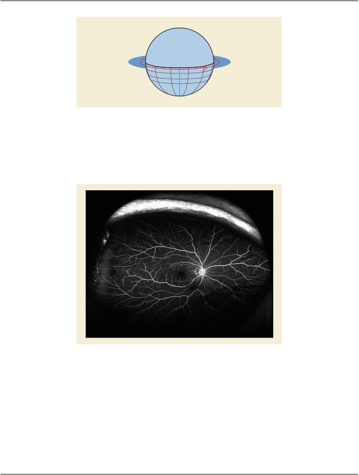
Certain projections are more suitable for a particular task than others. Conformal projections preserve angle, which is a property that​ applies to points in the plane of projection that are locally distortion-free. Practically speaking, this means that the projected meridian​ and parallel intersect through a point at right angles and are equiscaled. Therefore, measuring angles on the 2-dimensional image​ yields the same results as measuring these on the spherical representation, i.e., the retina. Conformal projections are particularly​ suitablefortaskswherethepreservationofshapesisimportant.Therefore,thestereographicprojectionexplainedinFigureUUU.1.2-​ 1 can be used for images on which to perform anatomically-correct measurements. The stereographic projection has the projection​ plane intersect with the equator of the eye where the fovea and cornea are poles. The points Fovea, p and q on the sphere (retina)​ are projected onto the projection plane (image in stereographic projection) along lines through the cornea where they intersect with​ the project plane creating points F′, p′and q′respectively.​

- Standard -​

DICOM PS3.17 2020a - Explanatory Information​

Page 715​

Fovea

p

p´ f

q´

q

Cornea

Figure UUU.1.2-1. Stereographic projection example​

Note that in the definition of stereographic projection the fovea is conceptually in the center of the image. For the mathematics below​ to work correctly, it is critical that each image is projected such that conceptually the fovea is in the center, even if the fovea is not in​ the image. This is not difficult to achieve as a similar result is achieved when creating a montage of fundus images; each image is​ re-projected relative to the area it covers on the retina. Most montages place the fovea in the center. An example of two images of​ the same eye in Figure UUU.1.2-2 and Figure UUU.1.2-3 taken from different angles and then transformed to adhere to this principle​ are in Figure UUU.1.2-4 and Figure UUU.1.2-5 respectively.​

Figure UUU.1.2-2. Image taken on-axis, i.e., centered on the fovea​

- Standard -​

Источник: https://studfile.net/preview/14585770/