Материал: part17

Внимание! Если размещение файла нарушает Ваши авторские права, то обязательно сообщите нам

DICOM PS3.17 2020a - Explanatory Information​

Page 951​

OOOO Encoding Perfusion Parameters for​ Parametric Maps and ROI Measurements​ (Informative)​

ThisAnnexcontainsexamplesofhowtoencodeperfusionmodelsandacquisitionparameterswithintheQuantityDefinitionSequence​ of Parametric Maps and in ROIs in Measurement Report SR Documents.​

Some measurements described as "relative" or "normalized" are calculated by dividing the measurement in a region of interest by a​ corresponding measurement from a reference region chosen for comparison purposes.​

The approach suggested is to describe that a perfusion value is being measured by using absolute or relative regional blood flow or​ volume (generic) as the concept name of the numeric measurement, and to add post-coordinated concept modifiers to describe:​

•​the general anatomic location and type of finding that mirror common usage of terminology specific to the application​

•​the size of any reference region used​

•​a coded description of the location of any reference region in terms of:​

•​anatomic site (drawn from CID 7192 "Anatomical Structure Segmentation Property Types")​

•​laterality (drawn from CID 244 “Laterality” or CID 246 “Relative Laterality” )​

Also illustrated is how the (121050, DCM, "Equivalent Meaning of Concept Name") can be used to communicate a single human​ readable textual description for the entire concept.​

The example used is from the oncology domain, but similar patterns can be used for other applications, e.g., for stroke.​

OOOO.1 Encoding Relative Cerebral Tumor Blood Flow for Parametric Maps​

This example shows how to use the Table C.7.6.16-12b “Real World Value Mapping Item Macro Attributes” in PS3.3 to describe pixel​ values of blood flow maps. It elaborates on the simple example provided in Section C.7.6.16.2.11.1.2 “Real World Values Mapping​ SequenceAttributes”byaddingcodedconceptsthatdescribethelocationofthemeasurementandthelocationandsizeofthereference​ region.​

•​Real World Value Mapping Sequence (0040,9096)​

•​...​

•​Real World Value Intercept (0040,9224) = "0"​

•​Real World Value Slope (0040,9225) = "1E-03"​

•​LUT Explanation (0028,3003) = "Relative Cerebral Tumor Blood Flow, relative to 150mm2 contralateral normal cerebellar gray​ matter"​

•​LUT Label (0040,9210) = "rCBF"​

•​Measurement Units Code Sequence (0040,08EA) = ({ratio}, UCUM, "ratio")​

•​Quantity Definition Sequence (0040,9220):​

•​CODE (246205007, SCT, "Quantity") = (126397, DCM, "Relative Regional Blood Flow)​

•​CODE (363698007, SCT, "Finding Site") = (12738006, SCT, "Brain")​

•​CODE (121071, DCM, "Finding") = (108369006, SCT, "Neoplasm")​

- Standard -​

Page 952​

DICOM PS3.17 2020a - Explanatory Information​

•​CODE (C94970, NCIt, "Reference Region") = (25991003, SCT, "Cerebellar Cortex")​

•​CODE (272741003, SCT, "Laterality") = (255209002, SCT, "Contralateral")​

•​NUMERIC (42798000, SCT, "Area") = 150 (mm2, UCUM, "mm2")​

•​TEXT (121050, DCM, "Equivalent Meaning of Concept Name") = "Relative cerebral tumor blood flow relative to 150mm2​ contralateral normal cerebellar gray matter"​

In this usage, the text of the (121050, DCM, "Equivalent Meaning of Concept Name") is redundant with the value of LUT Explanation​ (0028,3003); either or both could be omitted.​

The qualifiers of the quantity in this example, specifically the finding and finding site, are intentionally more generic (i.e., brain and​ neoplasm, respectively) than the more specific locations used in the SR examples that follow (i.e., left temporal lobe and primary​ neoplasm), since the purpose is to specify the type of the quantity, not encode an entire report in the quantity definition.​

Thenestingoftheindentedmodifiersintheexampleillustratesthatlateralityandareaaremodifiersofthereferenceregion(i.e.,encoded​ in the Content Item Modifier Sequence (0040,0441) of the Quantity Definition Sequence (0040,9220)).​

OOOO.2 Encoding Relative Cerebral Tumor Blood Volume for ROIs in Measure-​ ment Report SR Documents​

This example shows how to describe the total relative cerebral blood volume value of a region of interest that is a tumor in the left​ temporal lobe. In this case the template used is TID 1419 ROI Measurements within TID 1411 “Volumetric ROI Measurements” to​ separately encode the ROI with the total relative CBV and the ROI for the reference region with its size, with the relationship between​ them implicit in the presence of a coded reference region.​

For clarity, the enclosure of the content items within a Measurement Group container and the accompanying tracking identifiers and​ spatial information (coordinates and image and/or segmentation references) are not shown here.​

•​CODE (121071, DCM, "Finding") = (86049000, SCT, "Neoplasm, Primary")​

•​NUM (126398, DCM, "Relative Regional Blood Volume) = 1.2 ({ratio}, UCUM, "ratio")​

•​HAS CONCEPT MOD CODE (363698007, SCT, "Finding Site") = (78277001, SCT, "Temporal lobe")​

•​HAS CONCEPT MOD CODE (272741003, SCT, "Laterality") = (7771000, SCT, "Left")​

•​HAS CONCEPT MOD CODE (121401, DCM, "Derivation") = (255619001, SCT, "Total")​

•​HAS CONCEPT MOD TEXT (121050, DCM, "Equivalent Meaning of Concept Name") = "Total tumor blood volume relative to​ 150mm2 contralateral normal cerebral white matter"​

The reference region, its blood flow and size, would be specified as:​

•​CODE (121071, DCM, "Finding") = (C94970, NCIt, "Reference Region")​

•​NUM (126391, DCM, "Absolute Regional Blood Volume) = 34.6 (ml/(100.ml), UCUM, "ml/(100.ml)")​

•​HAS CONCEPT MOD CODE (363698007, SCT, "Finding Site") = (68523003, SCT, "Cerebral White Matter")​

•​HAS CONCEPT MOD CODE (272741003, SCT, "Laterality") = (255209002, SCT, "Contralateral")​

•​HAS CONCEPT MOD CODE (121401, DCM, "Derivation") = (255619001, SCT, "Total")​

•​NUM (42798000, SCT, "Area") = 150 (mm2, UCUM, "mm2")​

Alternatively, if the absolute blood volume of the reference region is not available, then its size can be specified alone, e.g.:​

•​CODE (121071, DCM, "Finding") = (C94970, NCIt, "Reference Region")​

•​NUM (42798000, SCT, "Area") = 150 (mm2, UCUM, "mm2")​

- Standard -​

DICOM PS3.17 2020a - Explanatory Information​

Page 953​

•​HAS CONCEPT MOD CODE (363698007, SCT, "Finding Site") = (68523003, SCT, "Cerebral White Matter")​

•​HAS CONCEPT MOD CODE (272741003, SCT, "Laterality") = (255209002, SCT, "Contralateral")​

The use of a specific reference region may be made explicit if the detailed information for both of the two source measurements, the​ absolute CBV for the lesion and the reference region, is available, in which case they could be encoded as their own instances of​ TID 1411 “Volumetric ROI Measurements”, and then the derived relative measurement encoded using TID 1420 "Measurements​ Derived From Multiple ROI Measurements", as follows:​

•​NUM (126398, DCM, "Relative Regional Blood Volume) = 1.2 ({ratio}, UCUM, "ratio")​

•​R-INFERRED FROM reference to measurement group content item of absolute measurement (Row 1 of TID 1411)​

•​R-INFERRED FROM reference to measurement group content item of reference region measurement (Row 1 of TID 1411)​

OOOO.5 Informative References​

This section lists useful references related to the taxonomy of perfusion measurements.​

OOOO.5.1 Perfusion Measurement Descriptions​

[Wetzel 2002] Radiology. Wetzel SG, Cha S, Johnson G, and et al. 2002. 224. 3. 797–803. “Relative Cerebral Blood Volume Meas-​ urements in Intracranial Mass Lesions: Interobserver and Intraobserver Reproducibility Study”. http://dx.doi.org/10.1148/​ radiol.2243011014 .​

[Bjørnerud 2010] J Cereb Blood Flow Metab. Bjørnerud A and Emblem KE. 2010. 30. 5. 1066–78. “A fully automated method for​ quantitative cerebral hemodynamic analysis using DSC–MRI”. http://dx.doi.org/10.1038/jcbfm.2010.4 .​

[Knutsson 2004] Magnetic Resonance Imaging. Knutsson L, Ståhlberg F, and Wirestam R. 2004. 22. 6. 789–98. “Aspects on the ac-​ curacyofcerebralperfusionparametersobtainedbydynamicsusceptibilitycontrastMRI:asimulationstudy”. http://dx.doi.org/​ 10.1016/j.mri.2003.12.002 .​

[Ziegelitz 2009] Magnetic Resonance in Medicine. Ziegelitz D, Starck G, Mikkelsen IK, and et al. 2009. 62. 1. 56–65. “Absolute​ quantification of cerebral blood flow in neurologically normal volunteers: Dynamic-susceptibility contrast MRI-perfusion​ compared with computed tomography (CT)-perfusion”. http://dx.doi.org/10.1002/mrm.21975 .​

[Jain 2011] AJNR. Jain R. 2011. 32. 9. 1570-1577. “Perfusion CT Imaging of Brain Tumors: An Overview”. http://doi.org/10.3174/​ ajnr.A2263 .​

[Wintermark2001]AJNR.WintermarkM,ThiranJP,MaederP,andetal.2001.22.5.905–14.“Simultaneousmeasurementofregional​ cerebral blood flow by perfusion CT and stable xenon CT: a validation study”. http://www.ajnr.org/content/22/5/905 .​

- Standard -​

Page 954​

DICOM PS3.17 2020a - Explanatory Information​

- Standard -​

DICOM PS3.17 2020a - Explanatory Information​

Page 955​

PPPP Real-Time Video Use Cases​ (Informative)​

PPPP.1 Introduction​

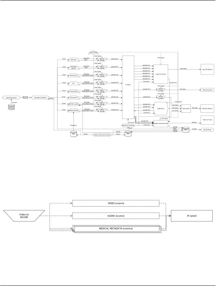
Figure PPPP.1-1. Overview diagram of operating room​

As shown in Figure PPPP.1-1, the DICOM Real-Time Video (DICOM-RTV) communication is used to connect various video or multi-​ frame sources to various destinations, through a standard IP switch, instead of using a video switch. In the future, the equipment​ producing video will support DICOM-RTV natively but it is anticipated that the first implementations will rely on the use of converters​ tocreateaDICOM-RTVstreamfromthevideostream(e.g.,SDI)andassociatedmetadatacomingfrominformationsystems,through​ existing mechanisms (e.g., DICOM Worklist). Such converters have to be synchronized with the Grand Master which is delivering a​ very precise universal time. Similarly, the video receivers (e.g., monitors) will be connected to the central switch via a converter which​ has also to be synchronized via the Grand Master. The different DICOM-RTV streams can be displayed, recorded, converted or​ combined together for different use cases. The medical metadata in the DICOM-RTV streams can be used to improve the quality of​ the whole system, as explained in the following use cases.​

Figure PPPP.1-2. Real-Time Video stream content overview​

As shown in Figure PPPP.1-2, the DICOM Real-Time Video stream is comprised of typically three different flows ("essences") for​ respectively video, audio and medical metadata information, using the intrinsic capability of IP to convey different flows on the same​ medium, multiplexing three kinds of blocks. There will be thousands of blocks for each video frame, hundreds for each audio sample​ and one for the medical metadata associated to each video frame, respectively represented as "V" (video) , "A" (audio) and "M"​ (metadata) on the Figure PPPP.1-3, which is the network view of the real-time streaming.​

- Standard -​

Источник: https://studfile.net/preview/14585770/