Материал: Oxford American Handbook of Urology ( PDFDrive )
BLADDER AND SPHINCTER BEHAVIOR 507
Pontine mesencephalic reticular formation is responsible for coordinating sphincter relaxation with detrusor contraction. Spinal cord lesions impair the transmission of coordinating influences from the pons during reflex detrusor contraction, and the uninhibited detrusor contraction stimulates a reflex sphincter contraction, resulting in bladder outflow obstruction.
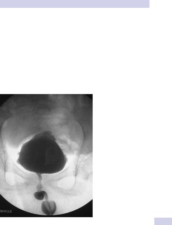
See Fig. 13.1. During electromyographic (EMG) recording, necessary to diagnose DSD, activity in the external sphincter increases during attempted voiding (the external sphincter should normally be “quiet” during voiding). An underactive sphincter is unable to maintain enough pressure, in the face of normal bladder pressures, to prevent leakage of urine.
Figure 13.1 Detrusor-external sphincter dyssynergia (DSD) seen during videocystourethrography.

508 CHAPTER 13 Neuropathic bladder
The neuropathic lower urinary tract: clinical consequences of storage and emptying problems
Neuropathic patients experience two broad categories of problems— bladder filling and emptying—depending on the balance between bladder and sphincter pressures during filling and emptying. The effects of these bladder filling and emptying problems include incontinence, retention, recurrent UTIs, and renal failure.
High-pressure sphincter
High-pressure bladder
If the bladder is overactive (detrusor hyperreflexia) or poorly compliant, bladder pressures during filling are high. The kidneys have to function against these chronically high pressures. Hydronephrosis can develop and ultimately cause renal failure.
At times the bladder pressure overcomes the sphincter pressure and the patient leaks urine (incontinence). If the sphincter pressure is higher than the bladder pressure during voiding, as in DSD, bladder emptying is inefficient (leading to retention, recurrent UTIs, or bladder calculi).
Low-pressure bladder
If the bladder is underactive (detrusor areflexia), pressure during filling is low. The bladder simply fills up—it is unable to generate enough pressure to empty causing retention and stasis of urine.
Urine leaks at times if the bladder pressure becomes higher than the sphincter pressure (incontinence), but this may occur only at very high bladder volumes or not at all.
Low-pressure sphincter
High-pressure bladder
If the detrusor is hyperreflexic or poorly compliant, the bladder will only be able to hold low volumes of urine before leaking (incontinence).
Low-pressure bladder
If the detrusor is areflexic, such that it cannot develop high pressures, the patient may be dry for much of the time. They may, however, leak urine (incontinence) when abdominal pressure rises (e.g., when coughing, rising from a seated position, or when transferring to or from a wheelchair). Their low bladder pressure may compromise bladder emptying, causing recurrent UTIs.
This page intentionally left blank

510 CHAPTER 13 Neuropathic bladder
Bladder management techniques for the neuropathic patient
A variety of techniques and procedures are used to treat retention, incontinence, recurrent UTIs, and hydronephrosis in the patient with a neuropathic bladder. A variety of neurological diseases, injury, congenital malformations, and medical conditions can result in a neuropathic bladder and variable degrees of voiding dysfunction. The incidence of voiding dysfunction with some common disorders is as follows:
•Cerebrovascular accident: 20–50%
•Parkinson disease: 35–70%
•Multiple sclerosis: 50–90%
•Diabetes mellitus; 5–59%
Each of the techniques described below can be used for a variety of clinical problems. Most patients require formal urodynamic evaluation (assessment of bladder filling, sensation, capacity, and compliance), urethral evaluation, voiding studies, and external sphincter electromyography to fully document the nature of the problem and identify the best clinical management.1
Thus, a patient with a high-pressure, hyperreflexic bladder that is causing incontinence can be managed with intermittent self-catheterization, sometimes supplemented with intravesical botulinum toxin injections, or a suprapubic catheter, or by sphincterotomy with condom sheath drainage, or by deafferentation combined with a sacral anterior root stimulator (SARS). Precisely which option to choose will depend on the individual patient’s clinical problem, their hand function, their lifestyle, resources available, and other personal factors, such as body image and sexual function.
Some patients will opt for a suprapubic catheter as a simple, generally safe, generally very convenient and effective form of bladder drainage. Others wish to be free of external appliances and devices because of an understandable desire to look and feel normal. They might opt for deafferentation with a SARS. The general principles of the management of the patient with neuropathic bladder dysfunction are as follows:
•Urodynamics are essential to plan urological management.
•Control intravesical pressure (prevent high pressure to protect upper tracts).
•Spontaneous voiding with continence is possible with NDO controlled medically.
•Urinary drainage: use intermittent catheterization or an external collection appliance.
•Indwelling catheterization is to be avoided long term because of complications (UTI, urethral erosion, calculi).
•Intermittent self-catheterization is the most effective treatment for most with detrusor areflexia, but requires low storage pressure.
1 Abrams P. Cardozo L. Fall M. et al. (2002). The standardization of terminology of lower urinary tract function: report from the Standardization Sub-committee of the International Continence Society. Neurourol Urodynam 21(2):167–178.

BLADDER MANAGEMENT TECHNIQUES 511
Medical therapy
Give anticholinergics to improve urinary storage pressure and decrease involuntary contraction. Use agents such as the following:
•Oxybutynin 5 mg PO tid–qid
•Hyoscyamine 0.125 mg PO qid
•Tolterodine LA 4 mg PO qd
A-Adrenergic blockers are used to decrease internal sphincter resistance and lower voiding pressure; they are ineffective for DSD. They may help control symptoms of autonomic dysreflexia. Medications include:
•Doxazosin 2 to 8 mg PO qd
•Terazosin 2 to 5 mg PO qd–bid
•Tamsulosin 0.4 mg PO qd
•Silodosin 8 mg PO qd
Botulinum toxin injection into the detrusor for detrusor overactivity is being used. The toxin selectively blocks acetylcholine release from nerve endings but is not FDA approved for bladder use.
Typical bladder regimens reported 10–30 individual endoscopic injections of 10 units/1 mL/site (posterior wall in midline, right and left lateral walls and dome), with sparing of the trigone.
Intermittent self-catheterization (ISC)
See p. 516.
Indwelling catheters, including suprapubic tubes
See p. 132.
External sphincterotomy
Deliberate ablation or stenting of the external sphincter is used to convert the high-pressure, poorly emptying bladder due to DSD to a low-pressure, efficiently emptying bladder. This is only for males with DSD; it requires a condom catheter afterward. Indications are retention, recurrent UTIs, recurrent bladder calculi, hydronephrosis, and renal insufficiency.
Techniques
•Surgical (with an electrically heated knife or vaporizing laser). Disadvantages are irreversibility, postoperative bleeding, septicemia, and stricture formation.2
•Intrasphincteric botulinum toxin. This technique is minimally invasive and reversible. Its disadvantage is that repeated injection is required every 3–9 months. It is not FDA approved for this use.
•Stenting (e.g., UroLume, AMS Minnetonka, MN). This is a 1.5–3 cm nonmetallic mesh alloy that expands to 42 F to keep the external sphincter open.3
2 JM Reynard (2003). Sphincterotomy and the treatment of detrusor–sphincter dyssynergia: current status, future prospects. Spinal Cord 41:1–11.
3 Chancellor M, Gajewski J, Ackman CFD, et al. (1999). Long-term follow-up of the North American Multicenter UroLume Trial for the treatment of external detrusor-sphincter dyssynergia. J Urol 161:1545–1550.