Материал: Oxford American Handbook of Urology ( PDFDrive )

Chapter 15 |
537 |
|
|
Pediatric urology
Embryology: urinary tract 538
Undescended testes 540
Urinary tract infection (UTI) 542
Vesicoureteric reflux (VUR) 544
Ectopic ureter 546
Ureterocele 548
Ureteropelvic junction (UPJ) obstruction 549
Hypospadias 550
Normal sexual differentiation 552
Abnormal sexual differentiation 554
Cystic kidney disease 558
Exstrophy 560
Epispadias 562
Posterior urethral valves 564
Non-neurogenic voiding dysfunction 566
Nocturnal enuresis 568

538 CHAPTER 15 Pediatric urology
Embryology: urinary tract
Following fertilization, a blastocyte (sphere of cells) is created, which implants into the uterine endometrium on day 6. The early embryonic disc of tissue develops a yolk sac and amniotic cavity, from which are derived ectoderm, endoderm, and mesoderm.
Organ formation occurs between 3 and 10 weeks’ gestation. Most of the genitourinary tract is derived from mesoderm.
Upper urinary tract
The pronephros (precursor kidney; pro = (Gk) before), derived from an intermediate plate of mesoderm, is present at weeks 1–4. It then regresses.
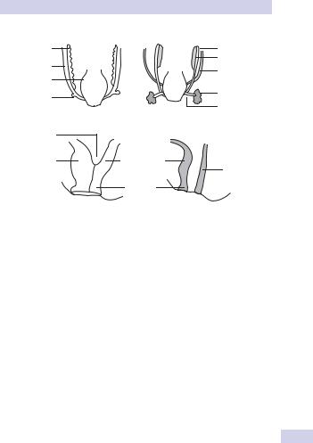
The mesonephros (meso = (Gk) middle) functions at weeks 4–8 and is also associated with two duct systems—the mesonephric duct and, adjacent to this, the paramesonephric duct (para = (Gk) beside) (Fig. 15.1).
The mesonephric (Wolffian) ducts develop laterally and advance downward to fuse with the primitive cloaca (hindgut). By week 5, a ureteric bud grows from the distal part of the mesonephric ducts and induces formation of the metanephros in the overlying mesoderm (permanent kidney; meta = (Gk) after).
Branching of the ureteric bud forms the renal pelvis, calyces, and collecting ducts. Glomeruli and nephrons are created from metanephric mesenchyme.
During weeks 6–10, the caudal end of the fetus grows rapidly and the fetal kidney effectively moves up the posterior abdominal wall to the lumbar region. Urine production starts at week 10.
Thus, in both males and females, the mesonephric duct forms the ureters and renal collecting system. The paramesonephric essentially forms the female genital system (fallopian tubes, uterus, upper vagina); in males, it regresses. The mesonephric duct forms the male genital duct system (epididymis, vas deferens, seminal vesicles, central zone of prostate); in the female, it regresses.
Lower urinary tract
The mesonephric ducts and ureters drain into the cloaca (Latin = sewer), which is later subdivided into the urogenital sinus (anteriorly) and the anorectal canal (posteriorly) during weeks 4–6 (see Fig. 15.1).
The bladder is formed by the upper part of the urogenital sinus. The lower part forms the urethra in females. In males, the mesonephric ducts form the posterior urethra, and closure of the urogenital groove creates the anterior urethra.

540 CHAPTER 15 Pediatric urology
Undescended testes
The testes descend into the scrotum in the third trimester (passing through the inguinal canal at 24–28 weeks). Failure of testicular descent results in cryptorchidism (or undescended testes).
Incidence
Incidence is 3% at birth (unilateral > bilateral). Approximately 80% will spontaneously descend by 3 months. The incidence at 1 year is 1%.
Classification
•Retractile: an intermittent active cremasteric reflex causes the testis to retract up and out of the scrotum.
•Ectopic (<5%): abnormal testis migration below the external ring of the inguinal canal (to perineum, base of penis, or femoral areas)
•Incomplete descent (~95%): testis may be intra-abdominal, inguinal, or prescrotal
•Atrophic/absent
Risk factors
These include preterm infants, low birth weight, small for gestational age, and twins.
Etiology
This includes abnormal testis or gubernaculum (tissue that guides the testis into the scrotum during development); endocrine abnormalities (low level of androgens, human chorionic gonadotrophin [hCG], luteinizing hormone (LH), calcitonin gene–related peptide); and decreased intraabdominal pressure (prune-belly syndrome, gastroschisis).
Pathology
There is degeneration of Sertoli cells, loss of Leydig cells, and atrophy and abnormal spermatogenesis.
Long-term complications
•Relative risk of cancer is 40-fold higher in the undescended testis. Most are seminomas; carcinoma in situ represents a small percentage (~2%). There is a slightly increased risk of cancer in the contralateral, normally descended testis.
•Reduced fertility
•Increased risk of testicular torsion
•Increased risk of direct inguinal hernias (due to a patent processus vaginalis)
Management
Full examination is required to elucidate if the testis is palpable and to identify location. Assess for associated congenital defects. If neither testis is palpable, consider chromosome analysis (to exclude an androgenized female) and hormone testing (high LH and FSH with a low testosterone indicates anorchia).

UNDESCENDED TESTES 541
Treatment should be performed within the first year. Hormone therapy (hCG, LHRH) stimulates testosterone production. Surgery consists of inguinal exploration, mobilization of spermatic cord, ligation of processus vaginalis, and securing the testis into a dartos pouch in the scrotal wall (orchidopexy). Laparoscopy can be used in planning surgery and for treatment.
Intra-abdominal testes may require division of spermatic vessels to provide extra length (Fowler-Stevens procedure, relying on collateral blood flow from vas), two-stage procedures, or microvascular autotransplantation.