Материал: Oxford American Handbook of Urology ( PDFDrive )

548 CHAPTER 15 Pediatric urology
Ureterocele
Definition
This is a cystic dilatation of the distal ureter as it drains into the bladder.
Incidence
Females are affected more than males (female–male ratio is 4:1). They predominantly affect Caucasians. 80% of cases are associated with the upper pole of a duplex system, although they can be found in single systems (more commonly in adults); 10% are bilateral.
Classification
•Intravesical: confined within the bladder
•Ectopic: if any part extends to the bladder neck or urethra
•Stenotic: intravesical ureterocele with a narrow opening
•Sphincteric: ectopic ureterocele with an orifice distal to the bladder neck
•Sphincterostenotic: orifice is both stenostic and distal to the bladder neck
•Cecoureterocele: ectopic ureterocele that extends into the urethra, but the orifice is in the bladder
Presentation
Infants commonly present with symptoms of UTI. Association with duplicated ureters increases the risk of reflux and reflux nephropathy.
Ureteroceles can also cause obstruction and hydronephrosis, which may be identified on antenatal ultrasound (US) scan or present in children with an abdominal mass or pain.
A prolapsing ureterocele can present as a vaginal mass in girls.
Investigation
•US shows a thin-walled cyst in the bladder often associated with a duplex system.
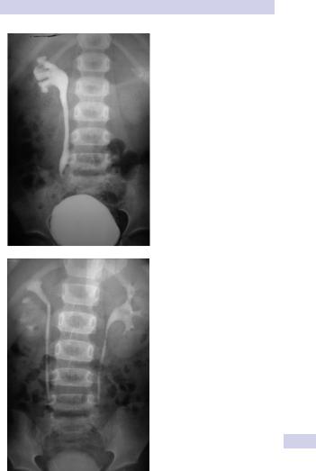
•Intravenous pyelogram (IVP) may demonstrate deviation of upperpole duplex kidney and delayed excretion of contrast signifying altered renal function. In single systems, contrast in the ureterocele gives the appearance of a cobra head.
•Voiding cystourethrogram can identify location, size, and associated reflux.
•Cystoscopy may reveal a defect near the trigone.
•99mTc-DMSA assesses renal segment function.
Treatment
Single-system ureterocele: Initial management is usually endoscopic incision of the ureterocele, which can be followed by surgical ureteric reimplantation to preserve renal function and prevent reflux.
Duplex-system ureterocele: Treatment options vary with the individual and include endoscopic incision, upper pole nephrectomy for a poorly functioning unit with ureterectomy (heminephroureterectomy), or, when there is useful renal function, ureteropyelostomy can be performed.

URETEROPELVIC JUNCTION (UPJ) OBSTRUCTION 549
Ureteropelvic junction (UPJ) obstruction
Definition
UPJ obstruction is a blockage of the ureter at the junction with the renal pelvis resulting in a restriction of urine flow.
Boys are affected more than girls. The left side is affected more often than the right side. Obstruction is bilateral in up to 40% of patients.
Etiology
In children, most UPJ obstruction is congenital, due to either an intrinsic narrowing (secondary to aberrant development of ureteric/renal pelvis muscle, abnormal collagen, or ureteral polyps) or extrinsic causes (compression of the UPJ by aberrant vessels).
Coexisting vesicoureteric reflux (VUR) is found in 40% of patients.
Presentation
UPJ obstruction is the most common cause of hydronephrosis found on prenatal and early postnatal US (differential diagnoses include UVJ obstruction, VUR, renal abnormalities, and posterior urethral valves). Infants may also present with an abdominal mass, UTI, and hematuria.
Older children present with flank or abdominal pain (exacerbated by diuresis), UTI, nausea and vomiting, and hematuria following minor trauma.
Investigation
If prenatal US has shown a large or bilateral hydronephrosis, a follow-up renal tract ultrasound scan should be performed soon after birth. If there is a prenatal unilateral hydronephrosis (and the bladder is normal), the scan is deferred until days 3–7 (to allow normal physiological diuresis to occur, which may spontaneously improve or resolve hydronephrosis).
If upper tract obstruction persists, a voiding cystourethrogram (VCUG) is indicated (to rule out VUR and examine for posterior urethral valves), and a renogram can assess individual renal function and drainage (DTPA, MAG-3).
Treatment
Children may be observed with US and renogram if they remain stable and have good renal function and no other complications (such as persistent infection or stones).
If children are symptomatic or have a significant hydronephrosis with impaired renal function (<40%), pyeloplasty is recommended.

550 CHAPTER 15 Pediatric urology
Hypospadias
Definition
Hypospadias is a congenital deformity in which the opening of the urethra (the meatus) occurs on the underside (ventral) part of the penis, anywhere from the glans to the perineum. It is often associated with a hooded foreskin and chordee (ventral curvature of the penile shaft).
It occurs in 1 in 250 live male births. There is an 8% incidence in offspring of an affected male, and a 14% risk in male siblings.
Classification
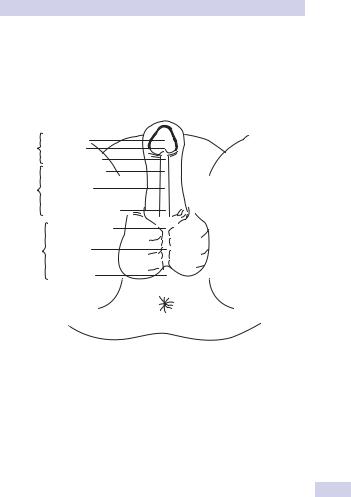
Hypospadias can be classified according to the anatomical location of the urethral meatus (Fig. 15.4).
•Anterior (or distal)—glandular, coronal, and subcoronal (~50%)
•Middle—distal penile, midshaft, and proximal penile (~30%)
•Posterior (or proximal)—penoscrotal, scrotal, and perineal (~20%)
Etiology
Hypospadias results from incomplete closure of urethral folds on the underside of the penis during embryological development. This is related to a defect in production or metabolism of fetal androgens, or the number and sensitivity of androgen receptors in the tissues.
Chordee is caused by abnormal urethral plate development, and the hooded foreskin is due to failed formation of the glandular urethra and fusion of the preputial folds (resulting in a lack of ventral foreskin but an excess of dorsal tissue).
Diagnosis
A full clinical examination will make the diagnosis. However, it is also important to seek out associated abnormalities that will need treatment (undescended testes, inguinal hernias, and hydroceles).
Patients with absent testes and severe hypospadias should undergo chromosomal and endocrine investigation to exclude intersex conditions.
Treatment
Surgery is indicated where deformity is severe, interferes with voiding, OR is predicted to interfere with sexual function. Surgery is now performed between 6 and 12 months of age. Local application of testosterone for 1 month preoperatively can help increase tissue size.
The aim of surgery is to correct penile curvature (orthoplasty), reconstruct a new urethra, and bring the new meatus to the tip of the glans using urethroplasy, glanuloplasty, and meatoplasty techniques.
Severe cases may require staged procedures. Common operations for anterior hypospadias include meatal advancement and glanuloplasty (MAGPI), meatal-based flaps (Mathieu procedure), and tubularization of the urethral plate.
Posterior defects require free grafts (buccal mucosa), on-lay grafts, and preputial transfer flaps.
HYPOSPADIAS 551
Complications
These include bleeding, infection, urethral strictures, meatal stenosis, urethrocutaneous fistula, urethral diverticulum, and failed procedures requiring reoperation.
Anterior Glanular
(Distal) Coronal Subcoronal
Distal penile
Middle Midshaft
Proximal penile
Penoscrotal
Posterior Scrotal (Proximal)
Perineal
Figure 15.4 The anatomical classification of hypospadias according to the location of the urethral meatus. This figure was published in Walsh PC, et al. Campbell’s Urology, 8th Edition, p. 2287. Copyright Elsevier 2002.