Материал: Oxford American Handbook of Urology ( PDFDrive )

542 CHAPTER 15 Pediatric urology
Urinary tract infection (UTI)
Definitions
UTI is a bacterial infection of the urine (>105 colony-forming units/ mL of urine), which may involve the bladder (cystitis) or kidney (pyelonephritis).
Classification
Children may be asymptomatic or symptomatic. It may be the first (initial) infection, recurrent UTI due to persistence of the causative organism and reinfection, or an unresolved infection due to inadequate treatment.
Incidence
Up to age 1 year, the incidence in boys is higher than in girls (males 2.7%: females 0.7%). Thereafter, the incidence in girls becomes greater (school age: males 1%; females 1–3%).
Pathology
Common bacterial pathogens are Escherichia coli (E. coli), Enterococcus, Pseudomonas, Klebsiella, Proteus, and Staphylococcus epidermis. Bacteria enter via the urethra to cause cystitis, and ascending infection causes pyelonephritis.
Alternatively, there can be hematogenous spread from other systemic infections.
Risk factors
•Age: Neonates and infants have increased bacterial colonization of the periurethral area and an immature immune system.
•Vesicoureteric reflux (VUR).
•Genitourinary abnormalities (UPJ obstruction; ureterocele; posterior urethral valves)
•Voiding dysfunction (abnormal bladder activity, compliance, or emptying).
•Gender (female > male after 1 year old)
•Foreskin: Uncircumcised boys have a 10-fold higher risk of UTI in the first year from bacterial colonization of the glans and foreskin.
•Fecal colonization (contributes to perineal bacterial colonization)
Presentation
UTI patients present with fever, irritability, vomiting, diarrhea, poor feeding, suprapubic pain, dysuria, voiding difficulties, incontinence, and flank pain.
Investigation
Diagnosis is made on urinalysis and culture. In young children, a catheterized urine specimen or a suprapubic aspirate is most accurate (bag specimens are less reliable because of skin flora contamination).
In toilet-trained children, a mid-stream specimen can be collected.

URINARY TRACT INFECTION (UTI) 543
Imaging
UTI in children <5 years, febrile UTI, infection in non–sexually active boys, and girls (>5 years) with two or more episodes of cystitis require renal tract imaging.
•Ultrasound identifies bladder and kidney abnormalities.
•Voiding cystourethrogram (VCUG) demonstrates urethral and bladder anomalies, VUR, and ureteroceles.
•DMSA (dimercaptosuccinic acid) renogram can demonstrate and monitor renal scarring.
Management
Empirical treatment should be started if infection is suspected. Children <3 months old with severe infection or pyelonephritis should receive broad-spectrum intravenous antibiotics (gentamicin and ampicillin) until antibiotic sensitivities are available.
Older children, and infants tolerating feeds can be given oral antibiotics (cephalosporins, or nitrofurantoin and trimethoprim-sulfamethoxide after 2 months old).
Complications
Neonates and young children have an increased risk of associated renal involvement and subsequent renal scarring, which can result in hypertension and renal failure.

544 CHAPTER 15 Pediatric urology
Vesicoureteric reflux (VUR)
Definition
VUR results from abnormal retrograde flow of urine from the bladder into the upper urinary tract.
Epidemiology
Overall incidence in children is >10%, with younger children affected more than older children, girls more than boys (female–male ratio 5:1). VUR occurs more often in Caucasian than in Afro-Caribbean children.
Siblings of an affected child have a 40% risk of reflux, and routine screening of siblings is recommended.
Pathogenesis
The ureter passes obliquely through the bladder wall (1–2 cm), where it is supported by muscular attachments that prevent urine reflux during bladder filling and voiding. The normal ratio of intramural ureteric length to ureteric diameter is 5:1.
Reflux occurs when the intramural length of ureter is too short (ratio <5:1). The degree of reflux is graded I–V (see Fig. 7.1, p. 345). The appearance of the ureteric orifice changes with increasing severity of reflux, classically described as stadium, horseshoe, golf-hole, or patulous.
Classification
•Primary reflux (1%) results from a congenital abnormality of the ureterovesical junction.
•Secondary reflux results from urinary tract dysfunction associated with elevated intravesical pressures. Causes include posterior urethral valves (reflux seen in 50%), urethral stenosis, neuropathic bladder, and detrusor sphincter dyssynergia (DSD).
•VUR is also seen with duplex ureters. The Weigert–Meyer rule states that the lower-pole ureter enters the bladder proximally and laterally, resulting in a shorter intramural tunnel, which predisposes to reflux.
Complications
VUR associated with UTI can result in reflux nephropathy with hypertension and progressive renal failure.
Presentation
Patients have symptoms of UTI, fever, dysuria, suprapubic or abdominal pain, failure to thrive, vomiting, and diarrhea.
Investigation
•Urinalysis and culture to diagnose UTI
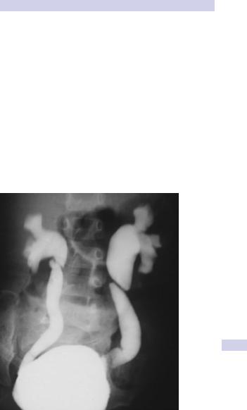
•Urinary tract ultrasound scan and VCUG to diagnose and grade reflux and establish reversible causes (Fig. 15.2)
•Urodynamic assessment if suspicious of voiding dysfunction
•DMSA scan to detect and monitor associated renal cortical scarring.

546 CHAPTER 15 Pediatric urology
Ectopic ureter
Definition
The ureteric orifice is situated below the normal anatomical insertion on the trigone of the bladder.
Pathogenesis
The ureteric bud arises from an abnormal position on the mesonephric duct during embryological development. Females are affected more than males (female–male ratio is 3:1). 80% of cases are associated with a duplicated collecting system (which predominantly affects females).
A duplex kidney has an upper pole and a lower pole, each with its own renal pelvis and ureter. The two ureters may join to form a single ureter, or they may pass down individually to the bladder (complete duplication). In this case, the upper pole ureter always opens onto the bladder below and medial to the lower pole ureter (Weigert–Meyer rule), predisposing to ectopic placement of the ureteric orifice.
Sites of ectopic ureters
•Females: bladder neck, urethra, vagina
•Males: posterior urethra, seminal vesicles, ejaculatory duct, vas deferens, epididymis, bladder neck
Presentation
Acute or recurrent UTI is common in both sexes. Obstruction of the ectopic ureter can lead to hydronephrosis and hydroureter, which may present as an abdominal mass.
•Females: When the ureteric opening is below the urethral sphincter, girls present with persistent vaginal discharge or incontinence, despite successful toilet training.
•Males: The ureter is always sited above the external urethral sphincter, so boys do not develop incontinence. UTIs may trigger epididymitis.
Investigation of urinary tract
•US demonstrates ureteric duplication, dilatation, and hydronephrosis.
•VCUG is used to assess reflux in lower pole ureters (Fig. 15.3).
•Cystourethroscopy can directly identify a ureteric opening in the urethra.
•Isotope renogram (99mTc-DMSA) is used to assess renal function to help plan surgery.
Treatment
An ectopic ureter is often associated with a poorly functioning renal upper pole or single-system kidney. In such cases, open or laparoscopic heminephrectomy or total nephrectomy with excision of the associated ureter is indicated.
When some function is retained in a single-system kidney, the distal ureter can be resected and reimplanted into the bladder.