Материал: Oxford American Handbook of Urology ( PDFDrive )

582 CHAPTER 16 Urological surgery and equipment
Patient safety in the operating room
It is a fundamental part of safe surgical practice to cross-check that the following have been done prior to starting an operation or procedure. The process of cross-checking should be done with another member of staff (time out).
•Patient identification. Confirm that you are operating on the right patient by a process of active identification (i.e., ask the patient their name, date of birth, and their address to confirm that you are talking to the correct patient).
•Ensure you are doing the correct procedure and on the correct side by cross-checking with the notes and X-rays. For lateralized procedures (e.g., nephrectomy, PCNL), the correct side of the
operation should be confirmed by cross-checking with the X-rays and with the X-ray report, as well as referring to the notes. Where it is possible for the sides of an IVP to be incorrectly labeled, this cannot happen with a CT scan, where the location of the liver (right side) and the spleen (left side) provide confirmation of what side is what. In addition, the patient should have the correct side mark on their body by the operating surgical team in the holding area.
•Check that appropriate antibiotic prophylaxis has been given.
•Check that DVT prophylaxis has been administered (e.g., heparin, AK-TEDS, intermittent pneumatic compression boots).
•Ensure that blood is available, if appropriate.
•The patient should be safely and securely positioned on the operating table—pressure points are padded, not touching metal (to avoid diathermy burns), body straps are securely in place.
Develop an approach to operating that involves members of your team. Listen to the opinions of staff members who are junior to you. They may sometimes be able to identify errors that are not obvious to you.
Cultivate the respect of the recovery room staff. They may express concern about a patient under their care—listen to their concerns, take them seriously, and, if all is well, reassure them.
It does no harm to your patients or to your reputation for you to develop the habit of visiting every patient in the recovery room in order to check that all is well. You may be able to identify a problem before it has developed into a crisis, and, at the very least, you will gain a reputation for being a caring surgeon.

TRANSURETHRAL RESECTION (TUR) SYNDROME 583
Transurethral resection (TUR) syndrome
TUR syndrome arises from the infusion of a large volume of hypotonic irrigating solution into the circulation during endoscopic procedures (e.g., TURP, TURBT, PCNL). It occurs in 0.5% of TURPs.
Pathophysiology
Biochemical, hemodynamic, and neurological disturbances occur:
•Dilutional hyponatremia is the most important—and serious—factor leading to the symptoms and signs. The serum sodium usually has to fall to <125 mmol/L before the patient becomes unwell.
•Hypertension is due to fluid overload.
•Visual disturbances may be due to the fact that glycine is a neurotransmitter in the retina.
Diagnosis—symptoms, signs, and tests
These include confusion, nausea, vomiting, hypertension, bradycardia and visual disturbances, and seizures. If the patient is awake (spinal anesthesia), they may report visual disturbances (e.g., flashing lights).
Preventing development of TUR syndrome and definitive treatment
Use a continuous irrigating cystoscope (provides low-pressure irrigation), limit resection time, avoid aggressive resection near the capsule, and reduce the height of the irrigant solution.1
For prolonged procedures, where a greater degree of fluid absorption may occur, measure serum Na and give 20–40 mg of IV furosemide to start off-loading the excess fluid that has been absorbed. If the serum sodium comes back as being normal, you will have done little harm by giving the furosemide, but if it comes back at <125mmol/L, you will have started treatment already and thereby may have prevented the development of severe TUR syndrome.
Techniques for measuring fluid overload (not commonly done)
•Weighing machines can be added to the ordinary operating table.2
•Adding a little alcohol to the irrigating fluid and constantly monitoring the expired air with a breathalyser3 allows an estimation of the volume of excess fluid that has been absorbed.
1 Madsen PO, Naber KG (1973). The importance of the pressure in the prostatic fossa and absorption of irrigating fluid during transurethral resection of the prostate. J Urol 109:446–452.
2 Coppinger SW, Lewis CA, Milroy EJG (1995). A method of measuring fluid balance during transurethral resection of the prostate. Br J Urol 76:66–72.
3 Hahn RG (1993). Ethanol monitoring of extravascular absorption of irrigating fluid. Br J Urol 72:766–769.

584 CHAPTER 16 Urological surgery and equipment
Catheters and drains in urological surgery
Catheters
Catheters are made from Latex or Silastic (for patients with Latex allergy or for long-term use—better tolerated by the urethral mucosa).
Types
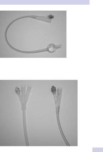
•Self-retaining (also known as a Foley, balloon, or two-way catheter) (Fig. 16.1). An inflation channel can be used to inflate and deflate a balloon at the end of the catheter, which prevents the catheter from falling out.
•Three-way catheter (also known as an irrigating catheter). Has a third channel (in addition to the balloon inflation and drainage channels) that allows fluid to be run into the bladder at the same time as it is drained from the bladder (Fig. 16.2).
Size
The size of a catheter is denoted by its circumference in millimeters. This is known as the “French” or “Charriere” (hence Fr) gauge. Thus a 12 Fr catheter has a circumference of 12 mm.
Uses
•Relief of obstruction (e.g., BOO due to BPE causing urinary retention—use the smallest catheter that you can pass; usually a 12 Fr or 14 Fr is sufficient in an adult)
•Irrigation of the bladder for clot retention (use a 20 Fr or 22 Fr threeway catheter)
•Drainage of urine to allow the bladder to heal if it has been opened (trauma or deliberately, as part of a surgical operation)
•Prevention of ureteric reflux, maintenance of a low bladder pressure, where the ureter has been stented (post-pyeloplasty for UPJ obstruction)
•To empty the bladder before an operation on the abdomen or pelvis (deflating the bladder gets it out of harm’s way)
•Monitoring of urine output postoperatively or in the critical patient
•For delivery of bladder instillations (e.g., intravesical chemotherapy or immunotherapy)
•To allow identification of the bladder neck during surgery (e.g., radical prostatectomy, operations on or around the bladder neck)
Drains
Drains are principally indicated for prevention of accumulation of urine, blood, lymph, or other fluids. They are particularly used after the urinary tract has been opened and closed by suture repair.
A suture line takes some days to become completely watertight, and during this time urine leaks from the closure site. A drain prevents accumulation of urine (a urinoma), the very presence of which can cause an ileus, and if it becomes infected, an abscess can develop.
CATHETERS AND DRAINS IN UROLOGICAL SURGERY 585
Figure 16.1 A Foley catheter with the balloon inflated.
Figure 16.2 Twoand three-way catheters.