Материал: Oxford American Handbook of Urology ( PDFDrive )

592 CHAPTER 16 Urological surgery and equipment
Irrigating fluids and techniques of bladder washout
Glycine is used for endoscopic surgery requiring application of diathermy. Normal saline is used for the following:
•Irrigation of bladder following TURP, TURBT
•Irrigation during ureteroscopy, PCNL
Blocked catheter post -TURP and clot retention
To avoid catheter blockage following TURP, keep the catheter bag empty; ensure a sufficient supply of irrigant solution.
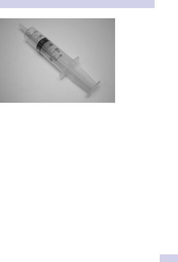
The bladder will be painfully distended. Irrigant flow will have stopped. A small clot may have blocked the catheter or a chip of prostate may have stuck in the eye of the catheter. Attach a bladder syringe to the end of the catheter and pull back (Fig. 16.9). This may suck out the clot or chip of prostate and flow may restart.
If it does not, draw some irrigant up into the syringe until it is about halffull and forcefully inject this fluid into the bladder. This may dislodge (and fragment) a clot that has stuck to the eye of the catheter.
If the problem persists, change the catheter. You may see the obstructing chip of prostate on the end of the catheter as it is withdrawn.
Blocked catheter post -TURBT
Use the same technique as for post-TURP catheter blockage, but avoid vigorous pressure on the syringe—the wall of the bladder will have been weakened at the site of tumor resection and it is possible to perforate the bladder, particularly in elderly women, who have thin bladder walls.
Blocked catheters following bladder augmentation or neobladder
The suture line of the augmented bladder is weak, and overvigorous bladder washouts can rupture the bladder.
IRRIGATING FLUIDS AND TECHNIQUES OF BLADDER WASHOUT 593
Figure 16.9 A bladder syringe—the tip is designed to fit onto a catheter.

594 CHAPTER 16 Urological surgery and equipment
JJ stents
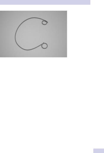
These are hollow tubes, with a coil at each end, which are inserted through the bladder (usually), into the ureter, and from there into the renal pelvis. They are designed to bypass a ureteric obstruction (e.g., due to a stone) or drain the kidney (e.g., after renal surgery). They have a coil at each end (hence the alternative name of “double pigtail” stent—the coils have the configuration of a pig’s tail—or the less accurate name of J stent).
JJ stents prevent migration downward (out of the ureter) or upward (into the ureter). They are therefore self-retaining.
They are made of polymers of variable strength and biodurability. Some stents have a hydrophilic coating that absorbs water and thereby makes them more slippery and easier to insert.
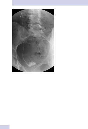
Stents are impregnated with bariumor bismuth-containing metallic salts to make them radio-opaque, so that they can be visualized radiographically to ensure correct positioning.
Types
Stents are classified by size and length. Common sizes are 6 Ch or 7 Ch (Fig. 16.10). Common lengths for adults are 22–28 cm. Multi-length stents are of variable length, allowing them to accommodate to ureters of different length.
Stent materials
These include polyurethane; silicone; C-flex; Silitek; Percuflex; and biodegradable material (the latter are experimental; they obviate the need for stent removal and eliminate the possibility of the “forgotten stent”).
Indications and uses
•Relief of obstruction from ureteric stones, benign (i.e., ischemic) ureteric strictures, or malignant ureteric strictures. The stent will relieve the pain caused by obstruction and reverse renal impairment if present.
•Prevention of obstruction post-ureteroscopy
•Passive dilatation of ureter prior to ureteroscopy
•To ensure antegrade flow of urine following surgery (e.g., pyeloplasty) or injury to ureter
•Following endopyelotomy (endopyelotomy stents have a tapered end from 14 Fr. to 7 Fr., to keep the incised ureter open)
Symptoms and complications of stents
•Stent symptoms that are common include suprapubic pain, LUTS (frequency, urgency—stent irritates trigone), hematuria, and inability to work.
•Urinary tract infection. Development of bacteriuria after stenting is common. In a small proportion of patients sepsis can develop. In such cases, consider placement of a urethral catheter to lower the pressure in the collecting system and prevent reflux of infected urine.
•Incorrect placement: too high (distal end of stent in ureter; subsequent stent removal requires ureteroscopy; can be technically difficult;
596 CHAPTER 16 Urological surgery and equipment
Figure 16.11 An encrusted stent.
Do stents cause obstruction?
In normal kidneys, stents cause a significant and substantial increase in intrarenal pressure that persists for up to 3 weeks.1 (This can be prevented by placing a urethral catheter.)
Do stents aid stone passage?
Ureteric peristalsis requires coaptation of the wall of the ureter proximal to the bolus of urine to be transmitted down the length of the ureter. JJ stents paralyze ureteric peristalsis. In dogs, the amplitude of each peristaltic wave (measured by an intraluminal ureteric balloon) falls (from 50 to 15 mmHg) and the frequency of ureteric peristalsis falls (from 11 to 3 waves per minute).
Peristalsis takes several weeks to recover; 3 mm ball bearings placed within a nonstented dog ureter take 7 days to pass, compared with 24 days in a stented ureter.
Are stents able to relieve obstruction due to extrinsic compression of a ureter?
Stents are less effective at relieving obstruction due to extrinsic obstruction by, for example, tumor or retroperitoneal obstruction.2 They are
1 Ramsay JW, et al. (1985). The effects of double J stenting on obstructed ureters. An experimental and clinical study. Br J Urol 57:630–634.
2 Docimo SG. (1989). High failure rate of indwelling ureteral stents in patients with extrinsic obstruction: experience at two institutions. J Urol 142:277–279.