Материал: Oxford American Handbook of Urology ( PDFDrive )

202 CHAPTER 6 Urological neoplasia
Prostate cancer: transrectal ultrasonography and biopsies
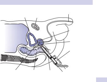
The most common diagnostic modality for prostate cancer is currently transrectal ultrasonography (TRUS) with guided biopsies (Fig. 6.1). TRUS provides imaging of the prostate and seminal vesicles using a 7.5 mHz biplane intrarectal probe measuring approximately 1.5 cm in diameter.
While TRUS imaging may be performed for other reasons (e.g., evaluation of infertility, sizing for treatment planning), a TRUS without biopsy is considered an inadequate way to evaluate for prostate cancer. Many advocate use of an enema before the biopsy to enhance visualization.
The ultrasound-guided periprostatic injection of local anesthetic (1% lidocaine) has become the standard in the United States when biopsy is planned. Rarely, general or regional anesthesia may be necessary, especially in the presence of anal stenosis or if saturation (>20) biopsies are planned.
A careful DRE precedes insertion of the probe. Broad-spectrum antimicrobials (e.g., quinolone antibiotic) are given before and after the procedure.
TRUS can image the outline of the prostate, cysts, abscesses, and calcifications within the prostate. Hypoechoic and hyperechoic lesions in the peripheral zone may be due to prostate cancer or inflammatory conditions, although most prostate cancers are in fact isoechoic and are not visualized on ultrasound.
Advanced biopsy techniques that use microbubble contrast agents with and without color Doppler are being actively investigated to improve yield. There are no convincing data that color Doppler alone can improve the yield of prostate biopsy over the standard grayscale biopsy.
Indications for TRUS without biopsy
•Accurate measurement of prostate volume for treatment planning (e.g., microwave, needle ablation, brachytherapy)
•Male infertility with azoospermia, to look for seminal vesicle and ejaculatory duct obstruction due to calculus or Müllerian cyst
•Suspected prostatic abscess (can be drained by needle aspiration)
•Investigation of chronic pelvic pain, looking for prostatic cyst or calculi
Indications for TRUS with biopsies
•An abnormal DRE and/or an elevated PSA (exceptions include very elderly men with massively elevated PSA and abnormal DRE, or those in whom a TURP is indicated for severe LUTS/retention where histology will be obtained). See Table 6.1 (p. 199).
•Previous biopsies showing multifocal high-grade PIN or ASAP
•Previous biopsies normal, but PSA rising or DRE abnormal
•To confirm viable prostate cancer following treatment if further treatment is being considered
PROSTATE CANCER: TRANSRECTAL ULTRASONOGRAPHY & BIOPSIES 203
|
Pubic |
Urethra |
Bladder |
symphysis |
|
|
|
Prostate |
Peritoneum |
|
gland |
|
|
|
|
|
Biopsy |
|
|
needle |
|
|
Spring-loaded |
|
|
biopsy needle |
|
|
‘gun’ |
Seminal |
Rectum |
Ultrasound |
vesicles |
|
probe |
Figure 6.1 Transrectal ultrasound scanning (TRUS). An ultrasound probe is inserted into the rectum to guide the biopsy needle into the correct position so that multiple core biopsies can be taken from different areas of the prostate.
Biopsy protocol
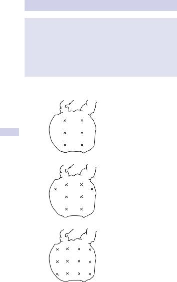
Ten to 12 18 French Tru-Cut needle biopsies are taken in a systematic fashion to include any palpable or sonographic target lesion. The traditional sextant protocol (a parasagittal base, mid-gland, and apex from each side) is no longer considered an adequate technique. To the 10–12 biopsy regimen, add samples from the far lateral peripheral zones (Fig. 6.2). Studies have demonstrated that these extra biopsies detect up to 15% more cancers.
Additional biopsies of each transition zone may be taken if a transition zone cancer is suspected or if a patient is undergoing repeat biopsies due to a rising PSA. Seminal vesicles biopsies occasionally add staging information if they appear abnormal on DRE, TRUS, or MRI.
Complications of prostatic biopsy
•Occasional vasovagal reaction (fainting) immediately after procedure
•Small risk of urinary tract infection and rarely urosepsis, which may be life threatening
•Small risk of significant rectal bleeding
•Mild hemospermia or hematuria, for several weeks after the procedure
Prostate cancer may also be diagnosed by transurethral resection of the prostate (TURP) histology or clinically (without histology) in certain circumstances. For example, it could be viewed as unnecessarily invasive to biopsy an elderly and frail symptomatic patient with a rock-hard prostate and a PSA of >100 ng/mL prior to commencing hormone therapy.
204 CHAPTER 6 Urological neoplasia
Box 6.2
It is not safe to biopsy a patient who is anticoagulated on warfarin; biopsying patients on low-dose aspirin remains controversial but is not considered unsafe by many. Other antiplatelet drugs (e.g., clopidogrel) are usually stopped for 7–10 days prior to biopsy.
It is important that the patient appreciates that negative biopsies do not exclude the possibility of prostate cancer, and that a positive result will not necessarily result in the recommendation of immediate treatment.
|
Seminal vesicles |
(a) |
|
|
Base |
Posterior |
Traditional |
surface of |
sextant |
prostate |
|
|
Apex |
(b)
8-core
(c)
12-core
Figure 6.2 Biopsy protocols. A 10to 12-core biopsy regimen is considered the standard of care today. Obtaining 4 apical cores can often be difficult.
This page intentionally left blank

206 CHAPTER 6 Urological neoplasia
Prostate cancer staging
Tumor staging uses the most current 2002 TNM (tumor, nodes, malignancy) classification (see Table 6.2 and Fig. 6.3). As with all cancer, prostate cancer staging may be considered clinical (prefixed with c) or pathological (prefixed with p), dependent on available data. (Note: In 2010 the American Joint Committee on Cancer (AJCC) will begin to incorporate PSA and Gleason Score into the staging system for prostate cancer.)
Tstage
Tstage is assessed primarily by digital rectal examination (see Fig. 6.3). Current imaging resolution limits reliability in detection of focal and microscopic extraprostatic extension of disease by means of MRI or TRUS. Recent prostatic biopsy may also confuse the interpretation of MRI images, particularly regarding the seminal vesicles.
Table 6.2 TNM (2002) staging of adenocarcinoma of the prostate
|
|
T0 |
No tumor (pT0 if no cancer found by histological examination) |
|
|
Tx |
T stage uncertain |
|
|
||
|
|
T1a |
Cancer nonpalpable on digital rectal examination (DRE), present |
|
|
|
in <5% of TURP specimens (in up to 18% of TURPs) |
|
|
T1b |
Cancer nonpalpable on DRE, present in >5% of TURP specimens |
|
|
T1c |
Cancer nonpalpable on DRE, present in needle biopsy taken |
|
|
|
because of elevated PSA |
|
|
T2a |
Palpable tumor, feels confined, in <half of one lobe on DRE |
|
|
T2b |
Palpable tumor, feels confined, in >half of one lobe on DRE |
|
|
T2c |
Palpable tumor, feels confined, in both lobes on DRE |
|
|
T3a |
Palpable tumor, locally advanced into periprostatic fat, unior |
|
|
|
bilateral and mobile on DRE |
|
|
T3b |
Palpable tumor, locally advanced into seminal vesicle(s) on DRE |
|
|
T4a |
Palpable tumor, locally advanced into adjacent structures, feels |
|
|
|
fixed on DRE |
|
|
T4b |
Palpable tumor, locally advanced into pelvic side-wall, feels fixed |
|
|
|
on DRE |
|
|
Nx |
Regional lymph not assessed |
|
|
N0 |
No regional lymph node metastasis |
|
|
N1 |
Tumor involves regional (pelvic) lymph nodes |
|
|
Mx |
Distant metastases not assessed |
|
|
M0 |
No distant metastasis |
|
|
M1a |
Nonregional lymph node metastasis |
|
|
M1b |
Tumor metastasis in bone |
|
|
M1c |
Tumor metastasis in other sites |
|
|
|
|